- Laboratory >
- Laboratory medicine >
- Cell imaging software
Cell imaging software
{{product.productLabel}} {{product.model}}
{{#if product.featureValues}}{{product.productPrice.formattedPrice}} {{#if product.productPrice.priceType === "PRICE_RANGE" }} - {{product.productPrice.formattedPriceMax}} {{/if}}
{{#each product.specData:i}}
{{name}}: {{value}}
{{#i!=(product.specData.length-1)}}
{{/end}}
{{/each}}
{{{product.idpText}}}
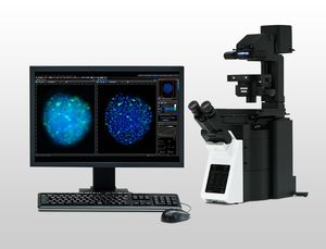
... Olympus cellSens platform gives you full control over the display and placement of icons, toolbars, and controls, enabling the software to grow and adapt to meet your evolving research needs. GET IN TOUCH cellSens ...
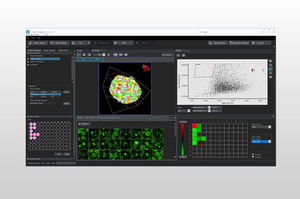
... Intelligent Answers NoviSight 3D cell analysis software advances your discovery by providing statistical data for spheroids and other 3D objects in microplate-based experiments. The software ...
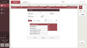
... and a User-friendly Interface User Friendly UI Software - Fast and easy patient registration - Fast procedure registration with preset positions - Positioning and imaging guide for accurate imaging - ...
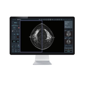
Advanced Image Parameter & Display Tools - Image Parameter Adjustment Tool : Easy image parameter setting tool for customized image style setting (3 default AIDIA optimized parameter provided) - Instant Image Compare : Compare before ...
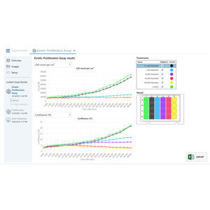
... lab. Cell Imaging Software — for and by cell biologists Cell imaging software can be overly complex and demanding ...
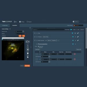
Inscoper Imaging Software is a turnkey hardware solution that completely revolutionizes the way fluorescence microscopes are controlled in live cell imaging. Today, ...

In addition to operating the VIVASCOPE hardware, the VIVASCAN software makes it easy to schedule patients for RCM exams, perform imaging exams on one or more lesions during a visit and review skin images ...

21 CFR part 11 compliant. Open platform communications (OPC) compliant. Visualize your cells: Monitor cell cultures in real time or live cell imaging. Cells ...

AmCAD-CA is a software device designed for analyzing digital cytological images. Considering morphology and chromatology, AmCAD-CA can be used to identify cytological features such as nuclear-cytoplasm ratio, anisokaryosis, ...

... analysis software developed for optical mapping and calcium imaging of the brain and heart. In addition to data files acquired with Brainvision's MiCAM imaging system, it is also possible ...

... processing algorithms. Areas of application include quantification of developed elispot plates, bacterial colonies, stained cells and tissues, gene and protein arrays. single click of a mouse it captures live ...

... it Works 1. Each cell is analyzed individually. Its number of sides, area, and perimeter are displayed in the cell table. 2. Mean and standard deviation are calculated for the selected sample. 3. ...
HAI Laboratories
Your suggestions for improvement:
the best suppliers
Subscribe to our newsletter
Receive monthly updates on this section.
Please refer to our Privacy Policy for details on how MedicalExpo processes your personal data.
- Brand list
- Manufacturer account
- Buyer account
- Our services
- Newsletter subscription
- About VirtualExpo Group


Please specify:
Help us improve:
remaining