- Laboratory >
- Laboratory medicine >
- Fluorescence cell imaging system
Fluorescence cell imaging systems
{{product.productLabel}} {{product.model}}
{{#if product.featureValues}}{{product.productPrice.formattedPrice}} {{#if product.productPrice.priceType === "PRICE_RANGE" }} - {{product.productPrice.formattedPriceMax}} {{/if}}
{{#each product.specData:i}}
{{name}}: {{value}}
{{#i!=(product.specData.length-1)}}
{{/end}}
{{/each}}
{{{product.idpText}}}
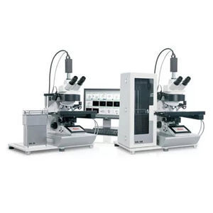
CytoVision is the one image analysis and management system that provides Cytogenetic laboratories with an integrated, scalable platform for brightfield and fluorescent samples, because only Leica combines ...
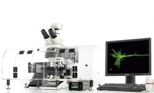
The LAS X Widefield Systems are ideal for applications in fluorescence microscopy and image analysis including live cell time-lapse experiments, multi-positioning, z-stacking and deconvolution. The ...
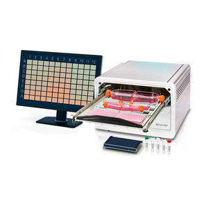
... Incucyte® S3 Live-Cell Analysis System, take your research further with automatic acquisition and analysis of cells in a physiologically relevant environment. See more information in ...
Sartorius Group
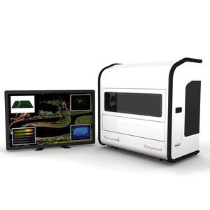
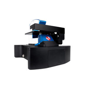
... pathology applications by combining confocal imaging with award-winning whole-slide scanning technology. Thanks to its innovative imaging technology, PANNORAMIC Confocal offers brightfield and fluorescence ...
... graph and green fluorescent cells Fluorescence Results in the Single Cell Tracking Assay Morphology screenshot from App Suite showing a dotplot and green fluorescent ...
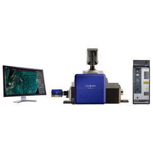
... Power Laser Engine (HLE) and new TIRF imaging modality, which exploits Borealis® illumination, B-TIRF (Borealis TIRF) for easy setup, more even illumination and thus more usable data ...
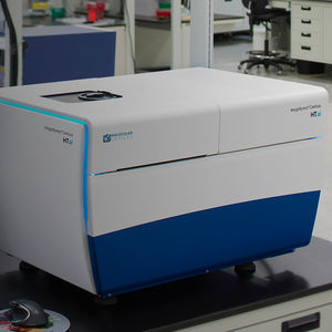
The ImageXpress® Confocal HT.ai High-Content Imaging System utilizes a seven-channel laser light source with eight imaging channels to enable highly multiplexed assays ...
Molecular Devices
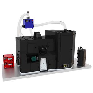
... inside an incubated environmental chamber. Automated imaging on a schedule The NIS-Elements Scheduler allows users to maintain their individual plates and schedule open times on the imaging system ...

... hard-coated fluorescence filters, and a computer with image analysis software. The sophisticated yet simple software supports multicolor fluorescence imaging, brightfield imaging, ...
Logos Biosystems
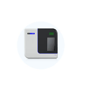
... software integrates the power of multispectral imaging with onboard spectral unmixing in a simplified workflow enabling spatial signatures at scale. Product Details As the premier and most highly cited imager ...
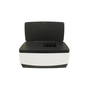
The VIVASCOPE 2500 is a confocal microscope specially designed for imaging fresh, needle aspirated, or fixed specimens in reflectance (phase contrast) mode or in fluorescence mode for specimens stained ...
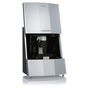
... physiological environment of cell cultures. Cell behavior and evaluation can be realized without staining. The system’s core technology uses high speed video imaging ...

Digital systems for microscopic analysis Biology and medicine — Cytology — Sperm analysis — Fluorescence — Pathology — Hematology Digital system for visualization ...
West Medica
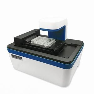
... live cell imaging system is designed to get time-lapse images and make taking cell videos much easier. KEY FEATURES - Compact and compatible with a standard CO₂ ...
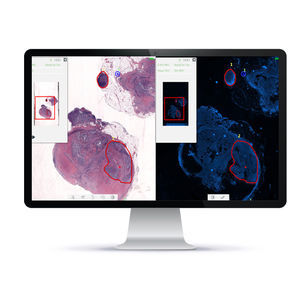
... Slide Imaging, digital FISH analysis and revolutionary digital tissue matching of FISH with H&E/IHC samples, PathFusion provides accurate and validated analysis for higher diagnostic ...
Applied Spectral Imaging

ntroducing a new, personal chemiluminescent imager that delivers high-quality, quantitative chemiluminescent and visible protein gel imaging through a unique web-based control software Why Do Scientists ...

... supports an array of applications and analyses. • - Assay your cells in brightfield and fluorescence – From label-free cell monitoring to fluorescence-based assays, ...
Axion BioSystems

... in FluorChem imagers ensure that you'll get the best image quality and the resolution you need to do accurate analysis, and differentiate between bands that are close together. The FluorChem M and R also enable multiplex ...
ProteinSimple

... Results Ultimate linearity for precise protein quantification over the full dynamic range. Multispectral Imaging Ultra-low noise imaging thanks to a dual camera amplifier architecture. Custom ...
Vilber GmbH
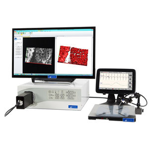
MRS CellLIVE is a powerful, handheld fluorescence confocal endomicroscope imaging system that is designed specifically for in vivo research of a variety of animal models ...
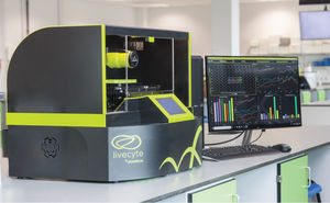
... Ptychographic quantitative phase imaging (QPI) technology for a range of label-free assays with or without up to seven channels of complementary fluorescence. Automated single-cell ...

Bringing High-Throughput Live Cell Imaging to Your Lab CELLCYTE X™ offers the user the ability to image live cells in real-time from within the incubator. From all of these images, ...

... and analog inputs recording functionality. Applications: - Widefield calcium imaging - Intrinsic optical signal (IOS) imaging - GCaMP imaging - FRET imaging Main ...
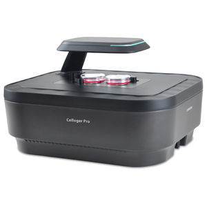
Celloger® Pro is an innovative live cell imaging system that redefines research capabilities. With its exceptional image quality and unmatched convenience, it empowers researchers with ...
Curiosis Inc

... easily detects GFP/RFP and other fluorescent markers in small animals. The capabilities of this in vivo imager enables research studies inclusive of whole animal down to individual cells. Focusing ...
Your suggestions for improvement:
the best suppliers
Subscribe to our newsletter
Receive monthly updates on this section.
Please refer to our Privacy Policy for details on how MedicalExpo processes your personal data.
- Brand list
- Manufacturer account
- Buyer account
- Our services
- Newsletter subscription
- About VirtualExpo Group


Please specify:
Help us improve:
remaining