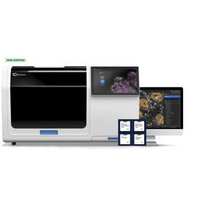
Automatic cell imaging system Xenium laboratorytissue3D

Add to favorites
Compare this product
Characteristics
- Operation
- automatic
- Applications
- laboratory, tissue
- Observation technique
- 3D
- Other characteristics
- high-resolution
Description
A state-of-the-art single cell spatial imaging platform
Seamlessly integrates transcript detection workflow, high-resolution imaging, decoding, and onboard data analysis.
Built to deliver exceptional image and data quality
Features state-of-the-art image acquisition and high-resolution optics with a nanometer-level localization precision.
Provides industry-leading throughput, up to 7x faster than other platforms
Generates best-in-class spatially resolved single cell gene expression data, at the fastest speeds per mm2. Analyze up to 236 mm2 per slide and up to 1,400 mm² per week.
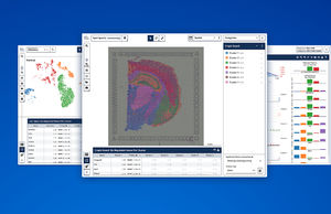
Converts terabytes of 3D imaging data into biologically meaningful insights
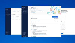
One-of-a-kind onboard analysis software performs high-powered computation on-instrument. Internal image sensor data is converted into ready-to-explore formats as soon your run finishes.
Delivers an unparalleled user experience
Simple run setup and cleanup operations, an intuitive touchscreen, and ergonomic design make instrument operation a breeze.
Expands with upcoming innovations
Flexible fluidic architecture allows the Xenium Analyzer to leverage new functionalities, assay enhancements, and more.
Dimensions (W x D x H)
52.5” x 27” x 31” (59” with door open)
133.3 cm x 68.5 cm x 78.7 cm (149.8 cm with door open)
Resolution
Transcript XY-localization precision < 30 nm and Z-localization precision< 100 nm
Pixel size = ~0.2 µm/pixel
Number of slides per run
2 slides
Throughput (area per time)
400 mm² in less than 50 hours
Total tissue area analyzable per run
~ 470 mm²
Sample prep
2–3 days sample prep, under 50 hour run time for 4 cm² of tissue on the Xenium Analyzer
Data output file formats
VIDEO
Catalogs
No catalogs are available for this product.
See all of 10xgenomics‘s catalogs*Prices are pre-tax. They exclude delivery charges and customs duties and do not include additional charges for installation or activation options. Prices are indicative only and may vary by country, with changes to the cost of raw materials and exchange rates.


