- Primary care
- General practice
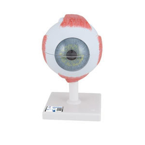
- Eye anatomical model
- 3B Scientific
Eye anatomical model F16for teaching
Add to favorites
Compare this product
Characteristics
- Area of the body
- eye
- Procedure
- for teaching
Description
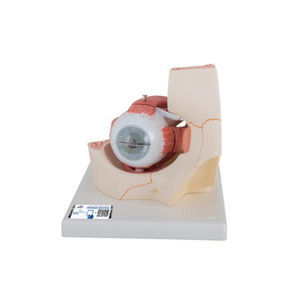
The MICROanatomy™ Eye model illustrates the microscopic anatomical structure of the retina with choroid and sclera. The left block-like, layered side of the eye model shows the complete structure of the retina including the supplying vascular layer and parts of the sclera from a light microscopic view.
The right part of the eye model is a sectional enlargement. MICROanatomy™ Eye shows the microscopic structure of the photoreceptors and the cells of the pigmented layer.
Catalogs
Exhibitions
Meet this supplier at the following exhibition(s):

Related Searches
- 3B Scientific anatomical model
- 3B Scientific training anatomical model
- 3B Scientific teaching anatomical model
- Stethoscope
- 3B Scientific bone model
- 3B Scientific skull model
- 3B Scientific flexible anatomical model
- Denture model
- 3B Scientific plastic anatomical model
- Oral anatomical model
- Vascular model
- 3B Scientific body anatomical model
- Training vascular model
- 3B Scientific spine anatomical model
- 3B Scientific leg anatomical model
- Cardiac anatomical model
- 3B Scientific pelvis model
- Digestive system model
- Circulatory system vascular model
- 3B Scientific nervous system model
*Prices are pre-tax. They exclude delivery charges and customs duties and do not include additional charges for installation or activation options. Prices are indicative only and may vary by country, with changes to the cost of raw materials and exchange rates.


