- Primary care
- General practice
- Tooth model
- 3B Scientific
Jaw anatomical model D26teethfor teaching
Add to favorites
Compare this product
Characteristics
- Area of the body
- jaw, teeth
- Procedure
- for teaching
Description
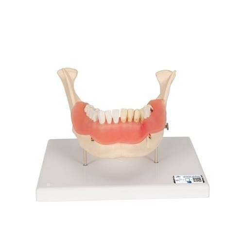
The dental disease model is based on a lifelike illustration of a lower jaw with 16 removable teeth of an adult magnified two times.
One half of the dental disease model shows eight healthy teeth and healthy gums.
The other half of the model shows the following dental diseases:
Dental plaque
Dental calculus (tartar)
Periodontitis
Inflammation of the root
Fissure, approximal and smooth surface caries.
One part of the front bone section can be removed from the dental disease model to view the roots, vessels and nerves. Two molars are sectioned along the length to show the inside of the tooth.
Catalogs
Exhibitions
Meet this supplier at the following exhibition(s):

Related Searches
- 3B Scientific anatomical model
- 3B Scientific training anatomical model
- 3B Scientific teaching anatomical model
- 3B Scientific bone model
- Stethoscope
- 3B Scientific flexible anatomical model
- 3B Scientific skull model
- Denture model
- 3B Scientific plastic anatomical model
- Oral anatomical model
- 3B Scientific body anatomical model
- Vascular model
- Training vascular model
- 3B Scientific spine anatomical model
- 3B Scientific leg anatomical model
- Cardiac anatomical model
- Digestive system model
- 3B Scientific pelvis model
- Circulatory system vascular model
- 3B Scientific nervous system model
*Prices are pre-tax. They exclude delivery charges and customs duties and do not include additional charges for installation or activation options. Prices are indicative only and may vary by country, with changes to the cost of raw materials and exchange rates.


