- Primary care
- General practice
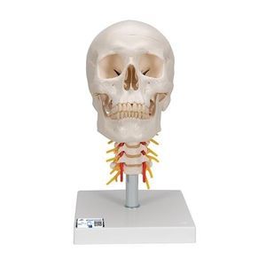
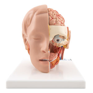
- Eye anatomical model
- 3B Scientific
Eye anatomical model F13for teaching
Add to favorites
Compare this product
Characteristics
- Area of the body
- eye
- Procedure
- for teaching
- Length
18 cm
(7.1 in)- Width
26 cm
(10.2 in)- Height
19 cm
(7.5 in)- Weight
1.4 kg
(3.09 lb)
Description
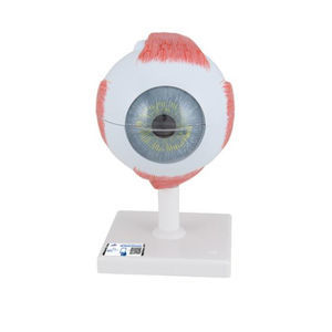
This large anatomical human eye model shows the optic nerve in its natural position in the bony orbit of the eye (floor and medial wall). At three times life size this eye model is great for anatomical demonstrations.
The human eyeball can be dissected into:
Both halves of sclera with cornea and eye muscle attachments
Both halves of the choroid with iris and retina
Eye lens
Vitreous humour
This high quality model is great for studying the anatomy of the human eye and the anatomy of the surrounding area! Human Eye Anatomy Model on base.
Every original 3B Scientific anatomy model now includes these additional FREE features:
Free access to the anatomy course 3B Smart Anatomy, hosted inside the award-winning
Complete Anatomy app by 3D4Medical
The 3B Smart Anatomy course includes 23 digital anatomy lectures, 117 different virtual
anatomy models and 39 anatomy quizzes to test your knowledge
Bonus: FREE warranty upgrade from 3 to 5 years with every product registration
TIP: You will also receive access to a free 3-day trial to all premium features of the Complete Anatomy app when you sign up for your 3B Smart Anatomy course.
Catalogs
Exhibitions
Meet this supplier at the following exhibition(s):

Related Searches
- 3B Scientific anatomical model
- 3B Scientific training anatomical model
- 3B Scientific teaching anatomical model
- Stethoscope
- 3B Scientific bone model
- 3B Scientific skull model
- 3B Scientific flexible anatomical model
- Denture model
- 3B Scientific plastic anatomical model
- Oral anatomical model
- Vascular model
- 3B Scientific body anatomical model
- Training vascular model
- 3B Scientific spine anatomical model
- Leg anatomy model
- Cardiac anatomical model
- 3B Scientific pelvis model
- Digestive system model
- 3B Scientific nervous system model
- Circulatory system vascular model
*Prices are pre-tax. They exclude delivery charges and customs duties and do not include additional charges for installation or activation options. Prices are indicative only and may vary by country, with changes to the cost of raw materials and exchange rates.


