
- Products
- Catalogs
- News & Trends
- Exhibitions
MRI test phantom DPHANTOMgeneral purpose
Add to favorites
Compare this product
fo_shop_gate_exact_title
Characteristics
- Type of calibration
- for MRI
- Area of the body
- general purpose
Description
IDENTIFYING IMAGE DISTORTIONS
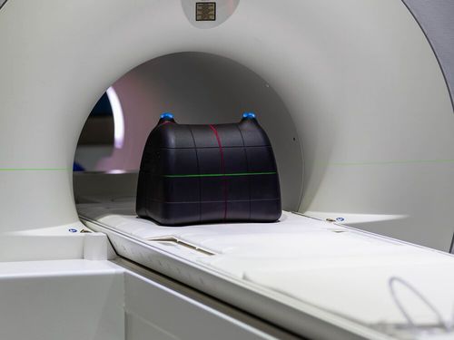
Magnetic Resonance Imaging (MRI) offers high soft-tissue contrast with significant potential utility within Radiation Oncology, particularly associated with delineation of targets and organs at risk, along with image guidance. It is prone however to geometric distortions related to both static field inhomogeneities and gradient field non-linearity. The dPhantom has been designed and created to facilitate high accuracy QC procedures associated with MRI Distortion. It may be filled with an appropriate fluid for MRI, as well as CT-imaged without fluid to facilitate image-registration assessment
DPHANTOM
3D One has produced a low cost, accurate Distortion phantom for the medical sector which we believe to be a superior alternative to traditional acrylic MRI Phantoms. The dPhantom is designed to assess and identify irregularities or distortions within magnetic resonance imaging scanners, commonly caused by the non-linear nature of the magnetic flux lines.
The unique leak-proof unibody nylon construction features a precise internal orthogonal 3-dimensional lattice. When imaged, the grid like structure provides excellent image contrast for effective distortion identification. The dPhantom can be filled with an appropriate fluid, such as nickel sulphate or nickel chloride, to optimise signal to noise and contrast.
Indexing grooves on the dPhantom’s exterior surface allow the device to be accurately positioned within the MRI scanner in all three orthogonal axes using the laser-alignment cross-hairs.
*Prices are pre-tax. They exclude delivery charges and customs duties and do not include additional charges for installation or activation options. Prices are indicative only and may vary by country, with changes to the cost of raw materials and exchange rates.
