- Secondary care
- Urology
- Urinary incontinence reconstruction mesh
- AMI - Agency for Medical Innovations
Urinary incontinence reconstruction mesh sensiTVTvaginal approachwomen
Add to favorites
Compare this product
Characteristics
- Mesh type
- urinary incontinence
- Surgical technique
- vaginal approach
- Patient type
- women
Description
The revolution in female sling surgery
A small design change can make a big difference
sensiTVT / sensiTVT-A – Adapts intra-operatively to patient’s anatomy
Traditional sling:
Tunnellers leave a smaller diameter tunnel in which cause traditional slings to fold! Nerves in the area may be compromised, potentially leading to so called “idiopathic sling failures” with symptoms such as discomfort, pain, voiding difficulties or de novo urge incontinence.
sensiTVT
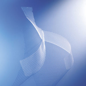
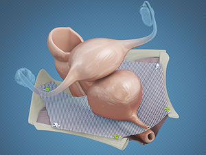
sensiTVT/sensiTVT-A adapts to the urethra due to passively articulating joints. It allows a parallel, flat mesh placement below and beside the urethra. This avoids overpressure zones caused by traditional slings because of the curled or twisted sling edges.
Twisted sensiTVT/sensiTVT-A – shows no effect below and beside the urethra
Pelvic floor ultrasound image in frontal plane B II – S – sensiTVT “buffer area” (two white points)
Pelvic floor ultrasound image in sagittal plane – sensiTVT/sensiTVT-A in optimal position. Good distance, flat and parallel, below the urethra
Ultrasound image in axial plane – sensiTVT in symmetric position
Adjustability:
sensiTVT-A is equipped with two groups of integrated sutures, which are left outside the skin following surgery, enabling optimal adjustment up to five days post-operatively. sensiTVT-A is especially indicated for patients after failed previous surgery, e.g. patients with low urethral mobility, patients with intrinsic sphincter deficiency (ISD) or obese patients.
Ultrasound imaging:
Dr. med. dr hab. J. Kociszewski, Hagen, Germany
Catalogs
sensiTVT
4 Pages
Other AMI - Agency for Medical Innovations products
Urogynaecology
Related Searches
- Reconstruction mesh
- Women reconstruction mesh
- Urinary incontinence reconstruction mesh
- Prolapse reconstruction mesh
- Vaginal approach reconstruction mesh
- Morcellator
- Cystocele reconstruction mesh
- Laparoscopic approach reconstruction mesh
- Uterine morcellator
- Colpocele reconstruction mesh
- Sphincter prosthesis
- Urinary incontinence sphincter prosthesis
- Vesical sphincter prosthesis
- Rectocele reconstruction mesh
- Hysterocele reconstruction mesh
*Prices are pre-tax. They exclude delivery charges and customs duties and do not include additional charges for installation or activation options. Prices are indicative only and may vary by country, with changes to the cost of raw materials and exchange rates.


