Cephalometric analysis software module 3D simulationfor dental imagingorthodontic

Add to favorites
Compare this product
Characteristics
- Function
- cephalometric analysis, 3D simulation
- Applications
- for dental imaging, orthodontic, surgical
Description
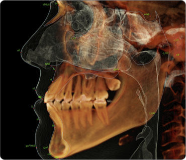
3D Analysis is the most advanced orthodontic analysis tool on the market. Utilizing unique 3D tracing methods, users can create more accurate and consistent tracings directly from the cone-beam CT scan. Many schools and researchers choose Anatomage’s 3D Analysis module as their main research and publication tool. Surgeons can quickly create surgical cuts and patient soft tissue simulations, making it a very powerful and accurate consultation tool. 3D Analysis can also be used for tracing standard 2D analyses, eliminating the need for reconstructing and tracing on ambiguous 2D X-ray images.
Comprehensive 3D Analysis
Visualize your cephalometric analyses in 3D space with our extensive and comprehensive landmark and measurement library or modify and create your own analysis.
Photo Face Wrap
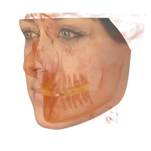
Superimpose a traditional clinical 2D photograph or a 3D facial image onto your patient’s scan to visualize soft tissue features and create the ultimate virtual patient
Impress Your Patients
Simulate soft-tissue movements with real-time orthognathic surgical cuts. Rotate and translate with accurate re-positioning tools to enhance your consultation.
ASSISTED LANDMARK IDENTIFICATION:
Anatomical landmarks have always been difficult to identify on flat cephalometric scans. To solve this problem, Anatomage created an innovative and simple way to perform cephalometric tracings in 3D that will automatically identify many specific points. Our comprehensive landmark and angle measurement library will allow you to accurately position points and easily define profiles on a 3D scan.
Simplify Cephalometric Tracings in 3D
Mathematically Accurate Landmark Identification
VIDEO
Catalogs
No catalogs are available for this product.
See all of Anatomage‘s catalogs*Prices are pre-tax. They exclude delivery charges and customs duties and do not include additional charges for installation or activation options. Prices are indicative only and may vary by country, with changes to the cost of raw materials and exchange rates.


