- Laboratory
- Laboratory medicine
- Automatic cell imaging system
- Applied Spectral Imaging
Automatic cell imaging system HiBand™laboratorydiagnosticcytogenetic

Add to favorites
Compare this product
Characteristics
- Operation
- automatic
- Applications
- laboratory, diagnostic, cytogenetic
- Observation technique
- fluorescence
Description
Artificial Intelligence based Chromosome Analysis and Karyotyping Solution
Digital Karyotyping | Automated Scanning
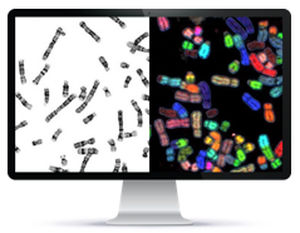
A state-of-the-art artificial intelligence (AI) system for digital chromosome analysis, featuring artificial-intelligence based automated workflow for high throughput karyotyping, as well as automated unattended scanning. HiBand provides significantly increased lab productivity and higher diagnostic confidence for Cytogenetic labs.
“HiBand doubled our lab productivity in Bone Marrow samples and tripled our productivity in Blood samples.”
Metaphases are detected in less than 2 minutes for entire slide.
Working in the background during image capture, automatic chromosome segmentation and karyotyping is completed.
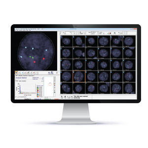
Successfully segmented and karyotyped metaphases are available for review and approval while scanning in a unique case gallery showing the original, enhanced and karyotyped image.
Metaphases are pre-sorted automatically by their spread, banding and overall quality.
Brightfield Karyotyping
G-Band and R-Band
Fluorescence Karyotyping
Q-Band, R-Band and FISH
Karyotype analysis requires minimal user adjustments.
Chromosomes are automatically segmented.
Count
Accurate automatic chromosome counting
Image Gallery
Display of all case metaphases and karyotypes
66% Technologist time savings!
Hours/Month
ully automated workflows for increased lab efficiencies
Start & walkaway scanning
Automatic barcode reading
Metaphase finding
Oil dispensing and smearing
Real-time image upload during slide scan
Export to lab LIS
Central Portal and Database | Easily Integrates with Lab LIS
Efficient
Comprehensive
Eliminates human error
VIDEO
Catalogs
No catalogs are available for this product.
See all of Applied Spectral Imaging‘s catalogsRelated Searches
- Automated cell imaging system
- Cell imager
- Laboratory cell imaging system
- Fluorescence cell imaging system
- Diagnostic cell imaging system
- Research cell imaging system
- Confocal cell imaging system
- Molecular biology cell imaging system
- Tissue cell imaging system
- 3D cell imaging system
- Pathology cell imaging system
- Clinical cell imaging system
- Cytogenetic cell imaging system
- FISH cell imaging system
- Spectral cell imaging system
*Prices are pre-tax. They exclude delivery charges and customs duties and do not include additional charges for installation or activation options. Prices are indicative only and may vary by country, with changes to the cost of raw materials and exchange rates.


