- Laboratory
- Laboratory medicine
- Histology reagent
- BIO-OPTICA Milano
- Company
- Products
- Catalogs
- News & Trends
- Exhibitions
Histology reagent 05-30010/Lliquid
Add to favorites
Compare this product
Characteristics
- Applications
- for histology
- Format
- liquid
Description
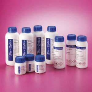
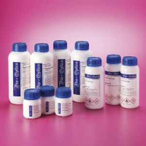
Primary container: white bottle in High Density Polyethylene (HDPE). Useful capacity 1 l. HDPE cap. Tamper evident cap.
Wear, water, alcohol and solvents resistant PVC label. Scratchproof ink resistant to water and alcohol.
Expected aim Product for the preparation of cyto-histological samples for optical microscopy.
Application Decolorizing solution used in Gram’s staining.
Principle
Two dyes are used one after the other: crystal violet and safranine. Crystal violet solution precipitates through oxidation with a iodine solution. The deriving complex attaches to bacteria cell walls with bonds of varying nature and intensity. The Gram's decolorizing solution removes the crystal violet-iodine complex from the walls of some bacteria, but it does not act on others. These retain the primary dye and are called Gram-positive. Decolorized bacteria are then counterstained with a red dye; they are called Gram-negative.
Method
For smears
1)Air dry the smear
2)Crystal violet solution according to Hucker 1 minute
3)Wash in distilled water
4)Gram’s iodine 1-2 minutes 5)Wash in distilled water6)Gram’s decolorizing solution 1 minute 7)Wash in distilled water
8)Safranine solution 1 minute
9)Wash in distilled water
10)Air dry the smear
11)Examine under the microscope in immersion oil
Results
Gram-positive bacteria..................................................................blue-violet
Gram-negative bacteria .................................................................red-orange
Storage Store the preparation at room temperature. Keep the containers tightly closed.
Catalogs
No catalogs are available for this product.
See all of BIO-OPTICA Milano‘s catalogsRelated Searches
- Bio-Optica solution reagent
- Laboratory reagent kit
- Bio-Optica histology reagent
- Reagent medium reagent kit
- Bio-Optica cytology reagent
- Bio-Optica stain reagent
- Buffer solution reagent kit
- Bacteria reagent kit
- Bio-Optica staining solution reagent
- Microscope slide
- Sample preparation reagent kit
- Pathology reagent
- Bilirubin reagent kit
- Bio-Optica fixative solution reagent
- Paraffin wax reagent
- Phosphate buffer reagent kit
- Microscopy reagent
- Collagen reagent kit
- Helicobacter pylori reagent kit
- Decalcifying solution reagent
*Prices are pre-tax. They exclude delivery charges and customs duties and do not include additional charges for installation or activation options. Prices are indicative only and may vary by country, with changes to the cost of raw materials and exchange rates.


