- Laboratory
- Laboratory medicine
- Staining solution reagent
- BIO-OPTICA Milano
- Company
- Products
- Catalogs
- News & Trends
- Exhibitions
Staining solution reagent Afogfor histologyfor cytology
Add to favorites
Compare this product
Characteristics
- Type
- staining solution
- Applications
- for histology, for cytology
Description
Minimum number of tests that can be performed 100
Completion time 22 minutes
Shelf life 2 years
Storage conditions 15-25°C
Additional equipment Not required
Application
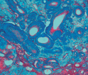
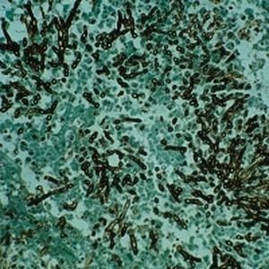
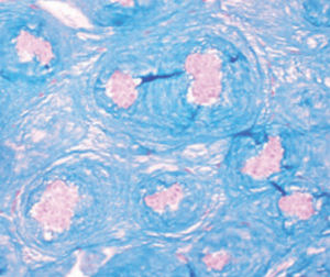
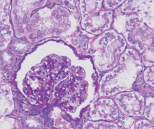
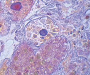
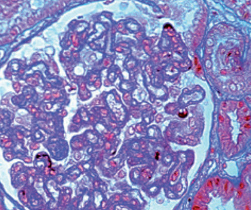
Reference method for highlighting protein deposits in renal biopsy.
Recommended fixative: Bouin.
Result
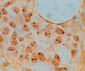
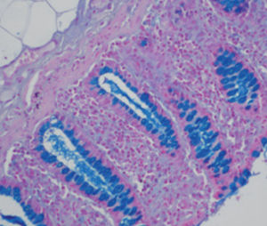
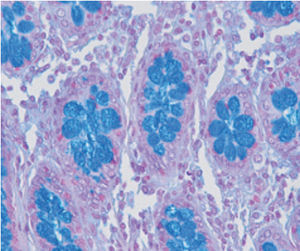
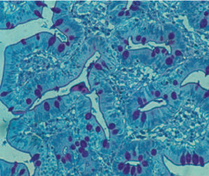
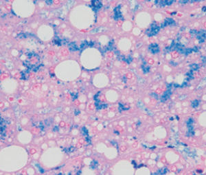
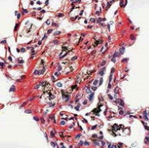
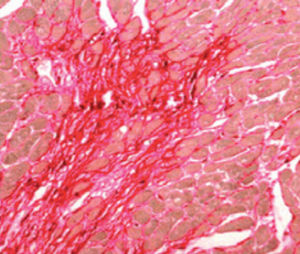
Collagen fibrils blue
Nuclei black
Erythrocytes, cytoplasm pink - orange
Elastic fibers pale pink - yellow or colorless
Protein deposits bright red
Product for the préparation of cyto-histological samples for optical microscopy.
Standard procedure for visualisation of glomerular protein deposits in kidney biopsy; recommended fixative: BOUIN
PRINCIPLE
In this method, three different dyes are used: Weigert hematoxylin for nuclear staining, orange G for cytoplasm and aniline blue for a sélective collagen staining.
Selectivity in this procedure is due to different degrees of affinity between dyes and tissue macromolecules. A central rôle is played by phosphomolibdic acid, which acts as a bound between tissue structures (collagen fibrils, cell membranes), and aniline blue (amphoteric dye). Orange G, which is the second component of AFOG solution, has no affinity to phosphomolibdic acid and is thus used to stain ail remaining structures. Acid fuchsin stains selectively glomerular protein deposits.
The product is intended for professional laboratory use for healthcare professionals.
Carefully read the information on the label (danger symbols, risk and safety phrases) and always consult the safety data sheet. Do not use if the primary container is damaged.
In the event of a serious accident, we recommended that you immediately inform Bio-Optica Milano S.p.A and the competent authorities.
Catalogs
General Catalogue
164 Pages
Related Searches
- Bio-Optica solution reagent
- Laboratory reagent kit
- Bio-Optica histology reagent
- Reagent medium reagent kit
- Bio-Optica cytology reagent
- Bio-Optica stain reagent
- Buffer solution reagent kit
- Bacteria reagent kit
- Bio-Optica staining solution reagent
- Microscope slide
- Sample preparation reagent kit
- Pathology reagent
- Bilirubin reagent kit
- Bio-Optica fixative solution reagent
- Paraffin wax reagent
- Phosphate buffer reagent kit
- Microscopy reagent
- Collagen reagent kit
- Helicobacter pylori reagent kit
- Decalcifying solution reagent
*Prices are pre-tax. They exclude delivery charges and customs duties and do not include additional charges for installation or activation options. Prices are indicative only and may vary by country, with changes to the cost of raw materials and exchange rates.


