- Laboratory
- Laboratory medicine
- Staining solution reagent
- BIO-OPTICA Milano
- Company
- Products
- Catalogs
- News & Trends
- Exhibitions
Staining solution reagent Gordon-Sweet for histologyfor cytology
Add to favorites
Compare this product
fo_shop_gate_exact_title
Characteristics
- Type
- staining solution
- Applications
- for histology, for cytology
Description
Minimum number of tests that can be performed 100
Completion time 40 minutes
Shelf life 1 year
Storage conditions 2-8°C
Additional equipment Not required
Application
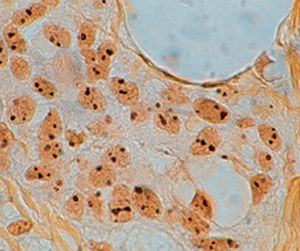
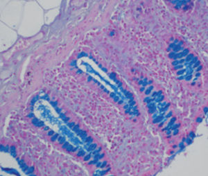
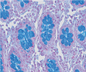
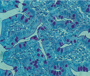
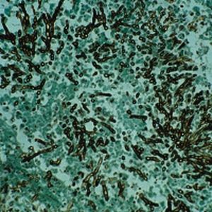
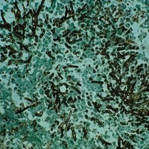
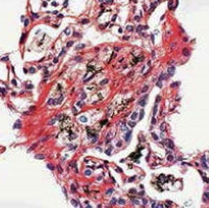
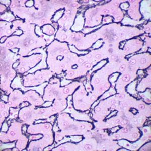
The method of choice for viewing argyrophilic reticular fibers of connective tissue.
Result
Reticular and nerve fibers black
Nuclei red, pink
Product for the preparation of cyto-histological samples for optical microscopy.
Recommended method to show argyrophilic reticular fibres in connective tissue.
PRINCIPLE
This method produces a selective evident impregnation in a very short time thanks to two factors: the preliminary impregnation
with an iron salt and the use as silver source of an unstable diaminic complex (ammoniacal solution), which is more reactive than
silver nitrate.
1) Pre-treatment with trivalent iron.
After a preparatory oxidation with potassium permanganate, the section is treated with trivalent iron (ferric ammonium
sulphate). Iron ions, more reactive than silver ions, quickly bind affine functional groups in argyrophilic structures.
2) Treatment with ammoniacal solution.
Silver is present in ammoniacal solution in the form of complex hydrosoluble oxide - [Ag(NH 3) 2] 2 O. This complex silver cation
replaces iron previously bound to tissues. In the next step, formic aldehyde acts as reducing agent: it removes oxygen from the
complex and releases metallic silver that deposits on argyrophilic structures.
[Ag(NH 3 )2 ]2 O + HCHO = 2 Ag + 4 NH 3 + HCOOH
Catalogs
General Catalogue
139 Pages
Related Searches
- Bio-Optica solution reagent
- Laboratory reagent kit
- Bio-Optica histology reagent
- Reagent medium reagent kit
- Bio-Optica stain reagent
- Bio-Optica cytology reagent
- Buffer solution reagent kit
- Bacteria reagent kit
- Bio-Optica staining solution reagent
- Microscope slide
- Sample preparation reagent kit
- Pathology reagent
- Bilirubin reagent kit
- Bio-Optica fixative solution reagent
- Paraffin wax reagent
- Phosphate buffer reagent kit
- Collagen reagent kit
- Helicobacter pylori reagent kit
- Microscopy reagent
- Decalcifying solution reagent
*Prices are pre-tax. They exclude delivery charges and customs duties and do not include additional charges for installation or activation options. Prices are indicative only and may vary by country, with changes to the cost of raw materials and exchange rates.


