- Laboratory
- Laboratory medicine
- Staining solution reagent
- BIO-OPTICA Milano
- Company
- Products
- Catalogs
- News & Trends
- Exhibitions
Staining solution reagent Gram for histologyfor cytology
Add to favorites
Compare this product
Characteristics
- Type
- staining solution
- Applications
- for histology, for cytology
Description
Minimum number of tests that can be performed 100
Completion time 40 minutes
Shelf life 2 years
Storage conditions 15-25°C
Additional equipment 3 vertical glass histology jars, funnel, filter, oven
Application
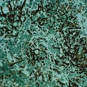
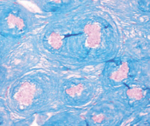
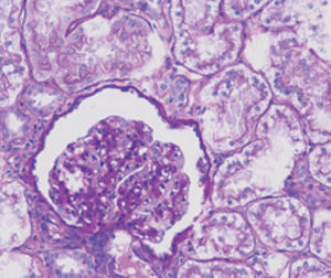
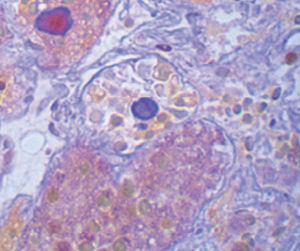
Method for differentiating Gram-positive and Gram-negative bacteria on histological
sections, smears and tissue apposition.
Result
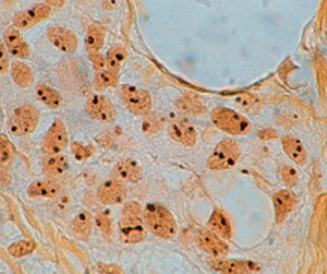
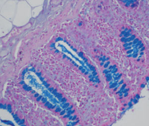
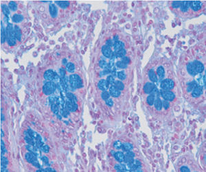
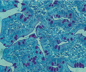
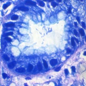
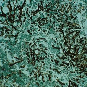
Gram-positive bacteria blue
Gram-negative bacteria red
Nuclei red
Product for the preparation of cyto-histological samples for optical microscopy.
To differentiate between Gram-positive and Gram-negative bacteria in tissue sections.
PRINCIPLE
Gram staining is the most important method to differentiate bacteria species. Two dyes are used one after the other: crystal
violet and fuchsine. Crystal violet solution precipitates through oxidation with a iodine solution. The deriving complex attaches
to bacteria cell walls with bonds of varying nature and intensity. The differentiating solution removes the crystal violet-iodine
complex from the walls of some bacteria, but it does not act on others. These retain the primary dye and are called Gram-
positive. Decolorized bacteria are then counterstained with a red dye; they are called Gram-negative. Gram-positive bacteria’s
capacity to retain the dye-iodine complex is usually ascribed to the bond which develops between the complex and a molecule
only Gram-positive possess, namely magnesium ribonucleate
Catalogs
General Catalogue
164 Pages
Related Searches
- Bio-Optica solution reagent
- Laboratory reagent kit
- Bio-Optica histology reagent
- Reagent medium reagent kit
- Bio-Optica cytology reagent
- Bio-Optica stain reagent
- Buffer solution reagent kit
- Bacteria reagent kit
- Bio-Optica staining solution reagent
- Microscope slide
- Sample preparation reagent kit
- Pathology reagent
- Bilirubin reagent kit
- Bio-Optica fixative solution reagent
- Paraffin wax reagent
- Phosphate buffer reagent kit
- Microscopy reagent
- Collagen reagent kit
- Helicobacter pylori reagent kit
- Decalcifying solution reagent
*Prices are pre-tax. They exclude delivery charges and customs duties and do not include additional charges for installation or activation options. Prices are indicative only and may vary by country, with changes to the cost of raw materials and exchange rates.


