- Laboratory
- Laboratory medicine
- Staining solution reagent
- BIO-OPTICA Milano
- Company
- Products
- Catalogs
- News & Trends
- Exhibitions
Staining solution reagent Grimelius for histologyfor cytology
Add to favorites
Compare this product
Characteristics
- Type
- staining solution
- Applications
- for histology, for cytology
Description
Minimum number of tests that can be performed 100
Completion time 3 hours 35 minutes
Shelf life 1 year
Storage conditions 2-8°C
Additional equipment Graduated cylinder, 50 ml histology jar, oven
Application
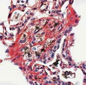
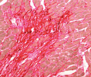
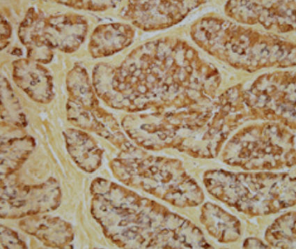
For viewing argyrophilic substances on tissue sections and appositions.
Result
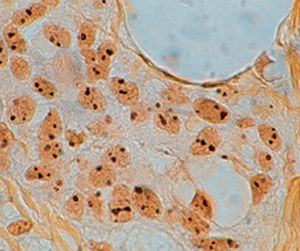
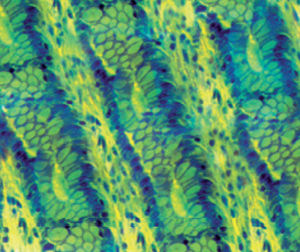
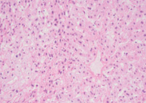
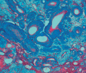
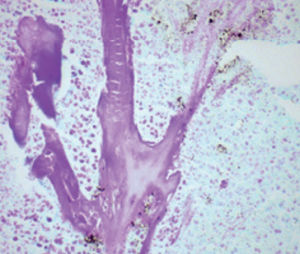
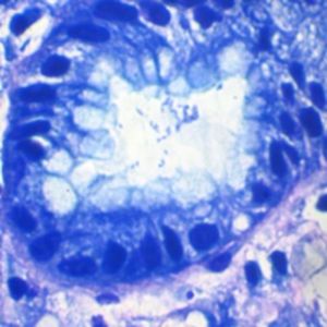
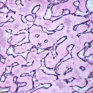
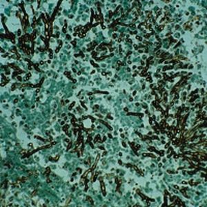
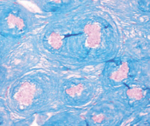
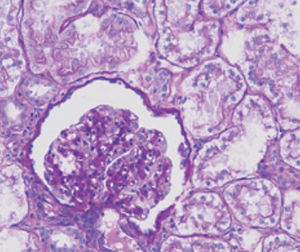
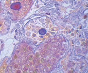
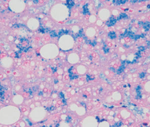
Argyrophilic granules from light brown to black
Product for the préparation of cyto-histological samples for optical microscopy. To show argyrophilic substances in tissue sections.
PRINCIPLE
This method is based on the intrinsic capacity of some tissue components to bind to silver halides. These are then revealed by a photographie process that reduces silver salts to metallic silver. Selectivity of method is due to a low concentration of silver salts in working solution. A second short imprégnation is used to precipitate more silver on the sites where it has already set down and therefore to make them blacker.
Method
1) Bring the section to the distilled water.
2) Dispense 10 drops of reagent A onto the section, leave to act for 40 minutes.
CAUTION: reagent A is corrosive. Handle with care and in an environment equipped
with an extractor fan. Avoid contact with the skin and eyes. Wear protective gloves
and goggles.
3) Wash twice in distilled water.
4) Dispense 10 drops of reagent B onto the section: leave to act for 10 minutes.
5) Without washing, drain the slide and dispense 10 drops of reagent C onto it: leave
to act for 2 minutes.
6) Without washing, drain the slide and dispense 10 drops of reagent D onto it: leave
to act for 3 minutes.
7) Wash in running tap water for 5 minutes.
8) Dehydrate by means of the ascending series of alcohols; xylene and balsam.
Catalogs
General Catalogue
164 Pages
Related Searches
- Bio-Optica solution reagent
- Laboratory reagent kit
- Bio-Optica histology reagent
- Reagent medium reagent kit
- Bio-Optica cytology reagent
- Bio-Optica stain reagent
- Buffer solution reagent kit
- Bacteria reagent kit
- Bio-Optica staining solution reagent
- Microscope slide
- Sample preparation reagent kit
- Pathology reagent
- Bilirubin reagent kit
- Bio-Optica fixative solution reagent
- Paraffin wax reagent
- Phosphate buffer reagent kit
- Microscopy reagent
- Collagen reagent kit
- Helicobacter pylori reagent kit
- Decalcifying solution reagent
*Prices are pre-tax. They exclude delivery charges and customs duties and do not include additional charges for installation or activation options. Prices are indicative only and may vary by country, with changes to the cost of raw materials and exchange rates.


