- Laboratory
- Laboratory medicine
- Staining solution reagent
- BIO-OPTICA Milano
- Company
- Products
- Catalogs
- News & Trends
- Exhibitions
Staining solution reagent Grocott MW for histologyfor cytology
Add to favorites
Compare this product
Characteristics
- Type
- staining solution
- Applications
- for histology, for cytology
Description
Minimum number of tests that can be performed 120
Completion time 50 minutes
Shelf life 1 year
Storage conditions 2-8°C
Additional equipment graduated cylinder, glass rod, oven
Application
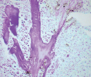
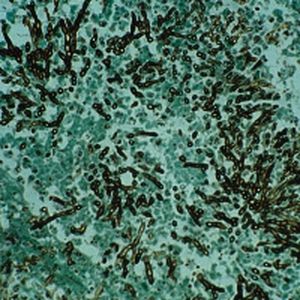
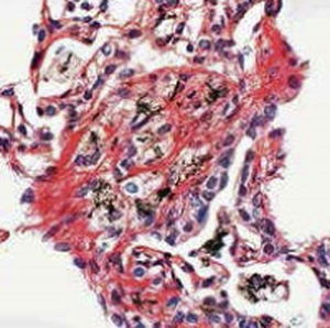
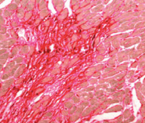
Method used for viewing fungi on a tissue section.
Result
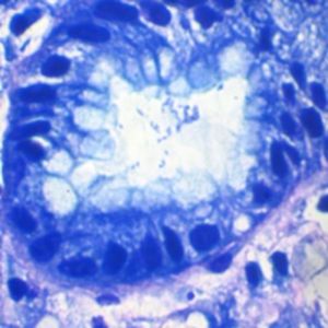
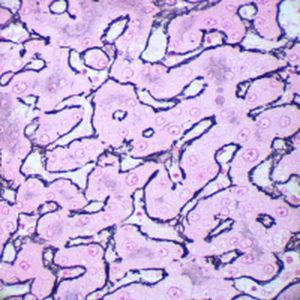
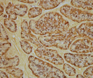
Fungi clearly outlined in black
Mucins dark gray
Background green
Product for the preparation of cyto-histological samples for optical microscopy.
To demonstrate fungi in tissue sections.
PRINCIPLE
In most fungi, cell walls are made of chitin, a polymer of N-acetylglucosamine together with polymers of D-glucose and D-mannose, proteins and lipids. Periodic acid reacts with glycolic and glycoaminic groups in polysaccharide chain oxidising them to
aldehydic groups and thus breaking the chain itself. These newly formed aldehydic groups reduce silver chloride, which is part of the silver-methenamine complex, to metallic silver and make it visible.
WARNING
For good results, follow these rules:
- Avoid to infect section and slide with no pathogenic fungi (handle only with gloves, do not expose preparation to air)
- Use always fresh distilled water.
- Use only clean glasswork.
- Avoid deposit of dust on section.
- Avoid contact between solutions and metal objects (tweezers...)
Catalogs
No catalogs are available for this product.
See all of BIO-OPTICA Milano‘s catalogsRelated Searches
- Bio-Optica solution reagent
- Laboratory reagent kit
- Bio-Optica histology reagent
- Reagent medium reagent kit
- Bio-Optica cytology reagent
- Bio-Optica stain reagent
- Buffer solution reagent kit
- Bacteria reagent kit
- Bio-Optica staining solution reagent
- Microscope slide
- Sample preparation reagent kit
- Pathology reagent
- Bilirubin reagent kit
- Bio-Optica fixative solution reagent
- Paraffin wax reagent
- Phosphate buffer reagent kit
- Microscopy reagent
- Collagen reagent kit
- Helicobacter pylori reagent kit
- Decalcifying solution reagent
*Prices are pre-tax. They exclude delivery charges and customs duties and do not include additional charges for installation or activation options. Prices are indicative only and may vary by country, with changes to the cost of raw materials and exchange rates.


