- Laboratory
- Laboratory medicine
- Staining solution reagent
- BIO-OPTICA Milano
- Company
- Products
- Catalogs
- News & Trends
- Exhibitions
Staining solution reagent Masson trichrome Goldner for histologyfor cytology
Add to favorites
Compare this product
Characteristics
- Type
- staining solution
- Applications
- for histology, for cytology
Description
Minimum number of tests that can be performed 100
Completion time 35 minutes
Shelf life 2 years
Storage conditions 15-25°C
Additional equipment Not required
Application
The method of choice for connective tissue, indicated for highlighting gametes, nuclei,
neurofibrils, glia, collagen, keratin, intracellular fibrils and negative images of the Golgi
apparatus.
Particularly indicated for black and white micro-photography.
Result
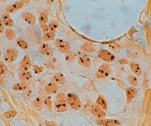
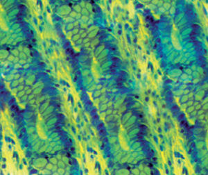
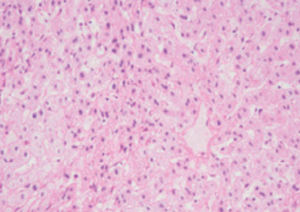
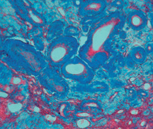
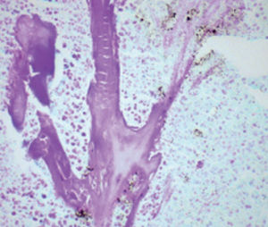
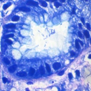
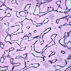
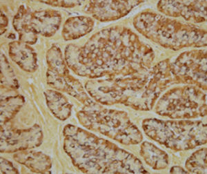
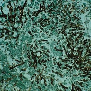
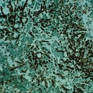
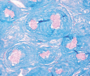
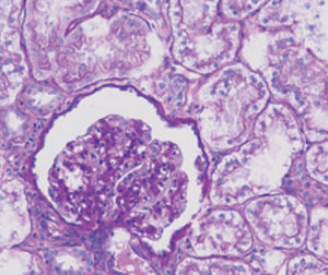
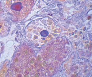
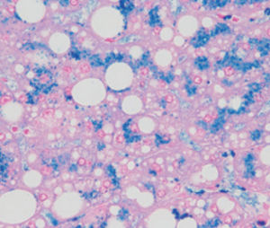
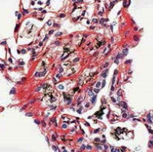
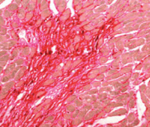
Nuclei and gametes black
Cytoplasm, keratin, muscle fibers, acidophilic granules red
Collagen, mucus, basophilic granules of the pituitary
gland
green
Delta cell granules of the pituitary gland green
Erythrocytes yellow
Product for the preparation of cyto-histological samples for optical microscopy.
Recommended method for connective tissue. It demonstrates gametes, nuclei, neurofibrils, neuroglia, collagen, keratin, intracellular fibrils, and negative image of Golgi apparatus. It is very suitable for black and white microphotographs.
PRINCIPLE
Four different stains are used: Weigert’s iron hematoxylin for nuclei, picric acid for erythrocytes, and a mixture of acid dyes (acid fuchsin-”ponceau de xylidine”) for cytoplasm and light green for collagen.
METHOD
1) Bring section to distilled water.
2) Put on the section 6 drops of reagent A and add 6 drops of reagent B: leave to act 10 minutes.
3) Without washing, drain the slide and put on the section 10 drops of reagent C: leave to act 4 minutes.
4) Wash quickly (3-4 seconds) in distilled water and put on the section 10 drops of reagent D, leave to act 4 minutes.
5) Wash in distilled water and put on the section 10 drops of reagent E: leave to act 10 minutes.
Catalogs
General Catalogue
164 Pages
*Prices are pre-tax. They exclude delivery charges and customs duties and do not include additional charges for installation or activation options. Prices are indicative only and may vary by country, with changes to the cost of raw materials and exchange rates.


