- Laboratory
- Laboratory medicine
- Staining solution reagent
- BIO-OPTICA Milano
- Company
- Products
- Catalogs
- News & Trends
- Exhibitions
Staining solution reagent 04-121812for histologyfor cytology
Add to favorites
Compare this product
Characteristics
- Type
- staining solution
- Applications
- for histology, for cytology
Description
Minimum number of tests that can
be performed
100
Completion time 45 minutes
Shelf life 2 years
Storage conditions 15-25°C
Additional equipment Not required
Application
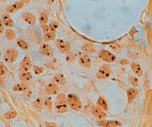
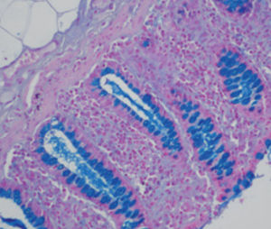
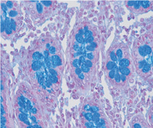
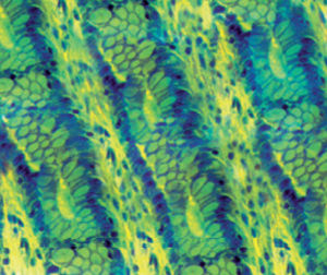
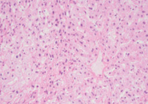
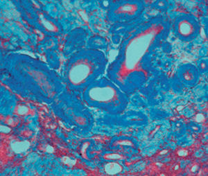
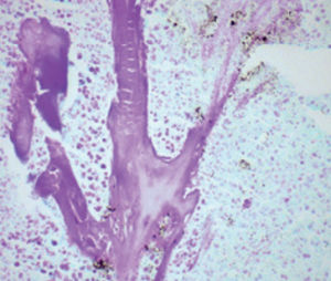
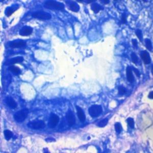
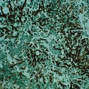
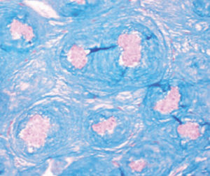
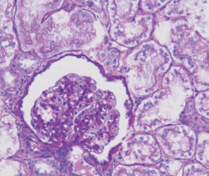
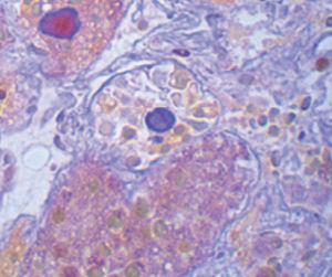
Method indicated for simultaneous viewing of DNA and RNA on histological sections.
Particularly indicated for highlighting plasma cells and RNA in histological sections and
cytological preparations.
Result
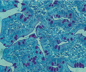
DNA pale green
RNA (plasma cells, nucleoli, blasts) pink – red
Mast cell granules blue
Background contrast turquoise
Product for the preparation of cyto-histological samples for optical microscopy.
To demonstrate simultaneously DNA and RNA in histologic sections. Recommended to show plasma cells and RNA in histologic
sections and cytological preparations.
PRINCIPLE
In this method, staining is obtained with a mixture of two basic dyes: purified methyl green and pyronin Y. In order to obtain a
differential stain, a buffer is added to the solution to reach pH 4,8. If pH is lower than 4,8 a red stain caused by pyronin prevails;
if pH is higher, a green-blue stain caused by methyl green prevails. The two dyes are not intrinsically affine to DNA and RNA; their
selectivity is a consequence of a definite pH value. This procedure may be difficult; we advise to read warning notes carefully
and to work in as a good conditions as possible.
Catalogs
General Catalogue
164 Pages
Related Searches
- Bio-Optica solution reagent
- Laboratory reagent kit
- Bio-Optica histology reagent
- Reagent medium reagent kit
- Bio-Optica cytology reagent
- Bio-Optica stain reagent
- Buffer solution reagent kit
- Bacteria reagent kit
- Bio-Optica staining solution reagent
- Microscope slide
- Sample preparation reagent kit
- Pathology reagent
- Bilirubin reagent kit
- Bio-Optica fixative solution reagent
- Paraffin wax reagent
- Microscopy reagent
- Phosphate buffer reagent kit
- Collagen reagent kit
- Helicobacter pylori reagent kit
- Decalcifying solution reagent
*Prices are pre-tax. They exclude delivery charges and customs duties and do not include additional charges for installation or activation options. Prices are indicative only and may vary by country, with changes to the cost of raw materials and exchange rates.


