- Surgery unit
- Surgical Instruments
- General surgery instrument kit
- BPB Medica - Biopsybell
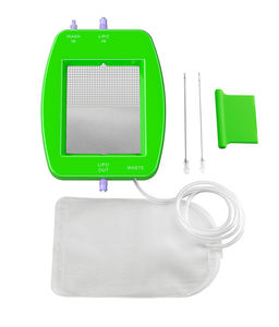
Bone marrow biopsy instrument kit ORTHOPLASTY™

Add to favorites
Compare this product
Characteristics
- Procedure
- bone marrow biopsy
Description
The subchondral bone plasty procedure is a minimally-invasive, fluoroscopically-assisted procedure that identifies and repairs subchondral bone defects, also named Bone Marrow Lesions (BMLs). The procedure is carried out with a minimally-invasive approach under fluoroscopy guidance along with arthroscopy, to target and manage findings inside the joint.
The pathology is classified as a SIFK (Subchondral Insufficiency Fracture of the Knee) and in the initial stages of SONK (Spontaneous Osteonecrosis of the Knee). The patient that presents with this pathology, suffers from relatively early osteoarthritis and consults the clinical specialist as a result of intense pain that does not correspond to a significantly compromised radiographic scenario.
In fact, these lesions are not visible under X-Ray and only a diagnostic confirmation using MRI reveals a hyper-intense uptake signal in sequences sensitive to T2 fluids (hydrogen) and in STIR sequences.
The objective of the method is to reinforce subchondral bone lesions using the same principle as vertebroplasty and involves the percutaneous insertion into the bone rarefaction site, of an appropriate bone substitute or of an autologous bone graft enhanced with a concentrate of mesenchymal stromal cells.
BENEFITS
Safe and precise minimally-invasive percutaneous approach
Fast procedure: approximately 20 minutes procedure
Rapid functional recovery
Pain relief after 1 day
Preservation of anatomical physiology for future operations
Reduced risk of infections
Ready-to-use bone substitute:
no preparation needed
Hardening in the wet environment only: no time pressure during application
VIDEO
Catalogs
No catalogs are available for this product.
See all of BPB Medica - Biopsybell‘s catalogs*Prices are pre-tax. They exclude delivery charges and customs duties and do not include additional charges for installation or activation options. Prices are indicative only and may vary by country, with changes to the cost of raw materials and exchange rates.


