- Laboratory
- Laboratory medicine
- Automatic cell imaging system
- Caliber Imaging & Diagnostics

- Products
- Catalogs
- News & Trends
- Exhibitions
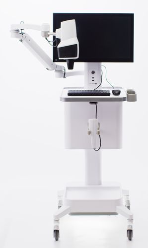
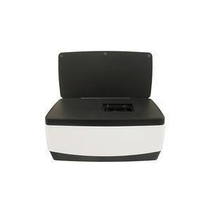
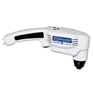
Automatic cell imaging system VivaScope® 1500laboratoryclinicalfor scientific research
Add to favorites
Compare this product
Characteristics
- Operation
- automatic
- Applications
- laboratory, clinical, for scientific research
- Cell type
- skin epithelial cells
- Observation technique
- confocal, in-vivo
- Other characteristics
- high-resolution
Description
The VIVASCOPE® system is an in vivo confocal imaging tool that uses a lowpowered laser to provide non-invasive, real-time, high-resolution images of the epidermis and the superficial collagen layers.* The system provides optical sectioning of unstained epithelium and the supporting stroma at a cellular level that may be reviewed by a physician to assist in forming a clinical judgment.
The VIVASCOPE 1500 reflectance confocal imaging system offers a non-invasive way to image the skin in vivo from the surface to the superficial collagen layers.*
Highlights
Capture up to 8 x 8 mm images
Macro and micro imaging
FDA 510(k) cleared**
Key Specifications
Mapped Field: 8 x 8 mm in both the X & Y directions Single Frame FOV: 500 µm x 500 µm
Displayed Image Resolution: 1024 x 1024 pixels
Depth of Imaging: Superficial collagen layers*
Image Formats: Native DICOM files exportable as: BMP, PNG, JPEG, and TIFF
CONFOCAL IMAGING
The VIVASCOPE® system is an in vivo confocal imaging tool that uses a lowpowered laser to provide non-invasive, real-time, high-resolution images of the epidermis and the superficial
collagen layers.* The system provides optical sectioning of unstained epithelium and the supporting stroma at a cellular level that may be reviewed by a physician to assist in forming a
clinical judgment.
VIVACAM DERMOSCOPY
VIVACAM® captures both clinical and dermoscopic images with a high-precision optical system.
High-resolution images Use to navigate within the macro image to specified corresponding confocal image
SINGLE IMAGE
The VIVASCOPE 1500 captures single images of skin that are parallel to the surface or on the “horizontal plane”.
Catalogs
VivaScope
4 Pages
VIVASCOPE® 3000
4 Pages
Other Caliber Imaging & Diagnostics products
CLINICAL IMAGING
Related Searches
- Microscopy
- Compound microscope
- Laboratory microscope
- Tabletop microscope
- Viewer software
- Laboratory software
- Online software
- Scan software
- Zoom microscope
- Research microscope
- Sharing software
- Automated cell imaging system
- Cell imager
- Interpretation software
- IR microscope
- Laboratory cell imaging system
- Confocal microscope
- Laser microscope
- Fluorescence cell imaging system
- Diagnostic cell imaging system
*Prices are pre-tax. They exclude delivery charges and customs duties and do not include additional charges for installation or activation options. Prices are indicative only and may vary by country, with changes to the cost of raw materials and exchange rates.


