- Laboratory
- Hematology
- Automatic hematology analyzer
- Cellavision AB
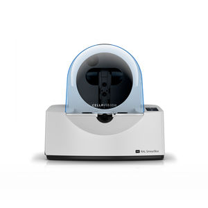
Automatic hematology analyzer DC-1benchtop100 samples

Add to favorites
Compare this product
Characteristics
- Operation
- automatic
- Configuration
- benchtop
- Loading capacity
- 100 samples
- Throughput
10 p/h
- Weight
11 kg
(24.3 lb)- Width
280 mm
(11 in)- Height
370 mm
(14.6 in)
Description
CellaVision DC-1 is the newest member of the CellaVision product family. It’s a smaller analyzer that has been custom-designed to enable low-volume hematology labs to implement CellaVisions digital methodology for performing blood cell differential.
Mode of operation
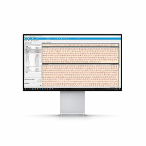
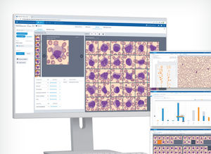
To perform a manual differential, a thin film of blood is wedged on a barcoded glass slide and stained according to the May-Grünwald Giemsa or Wright protocol. During slide processing the analyzer automatically locates, digitally captures and pre-classifies cells, after which the operator verifies and/ or modifies the suggested classification if necessary. The operator may also introduce additional observations and comments when needed.
Features
Automatically captures digital images of cells from blood smears
Creates digital scan of pre-defined area of any interesting specimen
SLIDE HANDLING
Accepts slides with ground edges, clipped, round or square corners.
Order ID for slides entered either manually or using an optional barcode reader.
Slides are loaded one slide at a time.
Analyzes slides with blood smears.
IMMERSION OIL
Manual oil dispensing
QUALITY CONTROL
Cell location accuracy test for the verification of the hardware and stain quality.
Built-in smear check.
ARCHIVING OF RESULTS AND IMAGES
Utilizing LAN
STORAGE CAPACITY
Secondary storage: Unlimited when transferred to external storage media.
PRINTER SUPPORT
Laser/ inkjet printers supported by Windows.
VIDEO
Catalogs
CellaVision®DC-1
12 Pages
*Prices are pre-tax. They exclude delivery charges and customs duties and do not include additional charges for installation or activation options. Prices are indicative only and may vary by country, with changes to the cost of raw materials and exchange rates.


