- Laboratory
- Laboratory medicine
- Staining solution reagent
- Celnovte Biotechnology Co., Ltd.

- Company
- Products
- Catalogs
- News & Trends
- Exhibitions
Staining solution reagent kit collagenfor histology
Add to favorites
Compare this product
Characteristics
- Type
- staining solution, collagen
- Applications
- for histology
Description
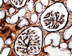
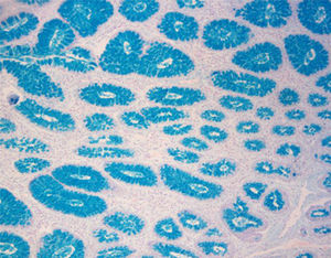
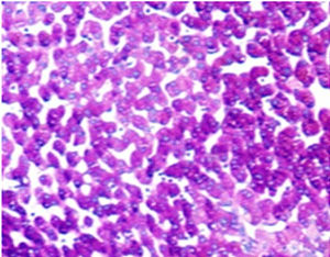
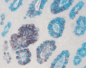
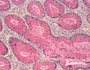
Reticular fibers are widely distributed and exist in two forms. One is a reticular scaffold for certain organs, such as bone marrow, spleen, lymph nodes, liver, thymus, and tonsils. The other is in the basement membrane of the epithelium. Meanwhile, smooth muscle, fat cells, capillaries, and nerve fibers are all covered with reticular fibers. There are no reticular fibers in the middle of the cancer tissue, but a large number of reticular fibros can be seen around its edge commonly known as cancer nests. Therefore, the reticular fibro staining is widely used in pathological diagnosis.
Product Features
Staining Principle
Reticular fiber, a special type of collagen, are difficult to identify with ordinary HE staining and show argyrophilia. The silver ammonia solution is absorbed by the tissue and binds to the protein in the tissue, reduced to black metal silver by formaldehyde and deposited in the tissue and on the surface. After toning with gold chloride, the unreduced silver salt is washed away with sodium thiosulfate solution to make the reticular fibers in the tissue clearly showed. It can comprehensively display the damage details of reticular scaffold in the pathological tissue.
Product Advantages
a)Clear organization structure, no obvious background staining
b)Stored for a long time
c)No precipitates and insoluble substances in each component reagent; Great stability
Catalogs
No catalogs are available for this product.
See all of Celnovte Biotechnology Co., Ltd.‘s catalogsOther Celnovte Biotechnology Co., Ltd. products
SPECIAL STAINS
Related Searches
- Assay kit
- Solution reagent kit
- Blood assay kit
- Molecular biology reagent kit
- Immunoassay assay kit
- Infectious disease detection kit
- Research reagent kit
- Protein reagent kit
- Diagnostic reagent kit
- Laboratory reagent kit
- Enzyme reagent kit
- Molecular test kit
- Histology reagent kit
- Clinical assay kit
- Reagent medium reagent kit
- Immunology reagent
- Cytology reagent kit
- Dye reagent
- Antibody
- Buffer solution reagent kit
*Prices are pre-tax. They exclude delivery charges and customs duties and do not include additional charges for installation or activation options. Prices are indicative only and may vary by country, with changes to the cost of raw materials and exchange rates.

