- Secondary care
- Ophthalmology
- Corneal topographer
- Chengdu SDK Medical Science and Technology

- Products
- Catalogs
- News & Trends
- Exhibitions
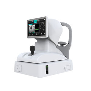
Corneal topographer sw-6000tabletop
Add to favorites
Compare this product
Characteristics
- Type of instrument
- corneal topographer
- Ergonomics
- tabletop
Description
The corneal topograph consists of 3 parts: ①Placido disc projection system: 28 or 34 rings are evenly projected onto the corneal surface from the center to the periphery, so that the entire cornea is within the analysis range of dry projection. ②Real-time image monitoring system; the ring image projected on the corneal surface can be observed, monitored and adjusted in real-time through the real-time image monitoring system, so that the corneal image can be taken in the best state, and then stored for analysis . ③Computer, image processing system; the computer first digitizes the stored images, applies the set calculation formulas and programs for analysis, and then displays the results of the analysis on the fluorescent screen with different color images. At the same time, the digitized statistical results are also together show.
The corneal topography is to analyze the entire corneal surface. Each projection ring has 256 points included in the processing system. Therefore, the entire cornea has more than 7000 data points entered into the analysis system. It can be seen that the corneal topography is systematic, accurate and precise. Corneal topography is clinically used to diagnose corneal astigmatism, quantitatively analyze corneal properties, and display corneal flexion in data or different colors. The difference between the two-axis flexion is corneal astigmatism. Diagnosing abnormal corneal flexion, the advent of the corneal map makes early diagnosis of keratoconus and keratoconus in the subclinical stage possible, and the accuracy of the diagnosis of keratoconus is as high as 96%.
Other Chengdu SDK Medical Science and Technology products
Eye Examination Equipment
Related Searches
- Ultrasound system
- B/W ultrasound system
- Tabletop laser
- Portable ultrasound system
- Fixed ophthalmic examination
- Tabletop ophthalmic examination instrument
- Ophthalmic biomicroscope
- Workstation with chair
- Hand-held ophthalmic examination instrument
- Table ophthalmic biomicroscope
- Ophthalmic workstation
- Ophthalmoscope
- Ophthalmic laser
- Refractometer ophthalmic examination
- Automatic refractometer
- Keratometer ophthalmic examination
- Automatic optical lens processing system
- Automatic keratometer
- Ophthalmic ultrasound imaging system
- Retinal camera
*Prices are pre-tax. They exclude delivery charges and customs duties and do not include additional charges for installation or activation options. Prices are indicative only and may vary by country, with changes to the cost of raw materials and exchange rates.


