Radiosurgery test phantom 603-GSfor MRIskull
Add to favorites
Compare this product
Characteristics
- Type of calibration
- for radiosurgery, for MRI
- Area of the body
- skull
Description
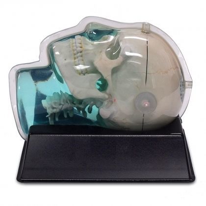
CIRS Model 603-GS was designed to assess MR image distortion in Stereotactic Radiosurgery Planning. It’s also a useful tool for verifying image fusion and deformable image registration algorithms used in various treatment planning systems. The tissue equivalent, anthropomorphic design closely matches a clinical imaging scenario. The phantom can be imaged using X-ray, Computed Tomography and Magnetic Resonance. It images well with all MRI sequences tested to date, including T1 weighted, T2 weighted, 3D Time of Flight, MPRAGE and CISS.
The skull is manufactured from a plastic-based trabecular bone substitute, and the interstitial and surrounding soft tissues are made from a proprietary signal generating water-based polymer. The entire phantom is encased in a clear plastic shell to protect gel from desiccation. It’s supplied with specially designed pads that allow fixation with any stereotactic frame or mounting devices for end-to-end testing. The phantom is also suitable for frameless SRS QA.
The entire inter-cranial portion of the skull volume is filled with an orthogonal 3D grid of 2.5mm diameter cross-like shaped rods spaced 10mm (I-S), 10.5mm (AP) and 11mm (L-R). Extra material added in the grid intersections increases grid signal. Five extended axis-rods intersect at the reference origin of the grid. The end of each extended axis is fitted with CT/MR markers allowing for accurate positioning with lasers and co-registration of CT and MR image sets.
The phantom includes right and left air voids, 3 mm in diameter by 17 mm long to simulate each ear canal for evaluation of potential distortions commonly found in clinical settings.
Catalogs
Related Searches
- Calibration phantom
- Tomography test phantom
- Radiography test phantom
- CT scan test phantom
- General purpose test phantom
- Ultrasound imaging test phantom
- Torso test phantom
- Head test phantom
- MRI test phantom
- Radiation therapy calibration phantom
- Breast test phantom
- Pediatric test phantom
- Abdomen test phantom
- Mammography test phantom
- Pelvis test phantom
- Whole body test phantom
- Skull test phantom
- Brain test phantom
- Fluoroscopy test phantom
- Lung test phantom
*Prices are pre-tax. They exclude delivery charges and customs duties and do not include additional charges for installation or activation options. Prices are indicative only and may vary by country, with changes to the cost of raw materials and exchange rates.


