- Laboratory
- Medical research
- Wire myograph
- Danish Myo Technology A.S (DMT)
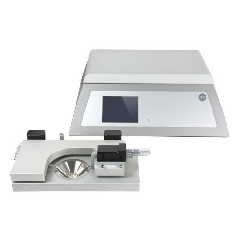
Wire myograph 360CWfor preclinical research
Add to favorites
Compare this product
Characteristics
- Type
- wire
- Application
- for preclinical research
Description
A perfect example are studies that correlate isometric contractions in an isolated, mounted blood vessel and intracellular Ca2+ measurements within the vascular smooth muscle cells.
The Confocal Wire Myograph System - 360CW is specially designed to provide very close optical access to the mounted artery or tissue segment, thereby allowing high-resolution images of fluorescent dyes or markers by laser scanning confocal microscopy (LSCM). This Wire Myograph system elegantly combines LSCM with artery myography to allow simultaneous measurements of isometric force and fluorescence imaging. A perfect example is studies that correlate isometric contractions in an isolated, mounted blood vessel and intracellular Ca2+ measurements within the vascular smooth muscle cells.
The chamber's unique design combines the precision and stability of conventional wire myographs with the added feature of precise Z-axis movement by a micrometer. This optimizes the flexibility of using this Wire Myograph System with different LSCMs and various high magnification objectives.
The bath design allows easy access to the high numerical aperture immersion objectives used on inverted microscopes and direct immersion objectives used on standard upright microscopes. Besides, special mounting supports were explicitly designed to allow precise vertical positioning of a mounted blood vessel or tissue ring directly above or on the chamber window. This permits the use of objectives with working distances smaller than 250μm on an inverted LSCM. This may be advantageous when simultaneous electrophysiological measurements are collected from the top of the mounted tissue.
Catalogs
360CW
2 Pages
WIRE MYOGRAPH SYSTEMS
4 Pages
Other Danish Myo Technology A.S (DMT) products
Wire Myograph Systems
Related Searches
*Prices are pre-tax. They exclude delivery charges and customs duties and do not include additional charges for installation or activation options. Prices are indicative only and may vary by country, with changes to the cost of raw materials and exchange rates.


