- Primary care
- General practice
- Mandible model
- Denoyer-Geppert

- Products
- Catalogs
- News & Trends
- Exhibitions
Bone model 0106-00mandiblemandibularjaw
Add to favorites
Compare this product
fo_shop_gate_exact_title
Characteristics
- Area of the body
- bone, mandible, mandibular, jaw, head, hand, neck, teeth, side, blood vessels
- Type
- canine
- Material
- plastic
- Length
14 in
- Width
9 in
- Height
4 in
Description
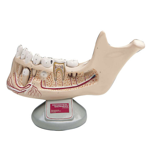
The lower jaw of an eleven year old depicts deciduous teeth being replaced by permanent teeth. Unbreakable plastic, the model has been scaled to 3-1/2 times life size to enhance difficult to observe details.
The outer portion of the jaw bone has been cut away exposing the roots of the teeth, their nerves and blood vessels. Four teeth can be extracted from the jaw: an incisor, a premolar, molar, and a developing canine. One molar has caries. Another is sectioned longitudinally revealing its enamel, dentine, cementum, and pulp.
Thirty-one hand numbered structures are identified in the accompanying key.
The Teeth
(Teeth shown would be for a 10-12 yr. old. Development and eruption ages vary greatly between individuals.)
(Left side of mandible shown on model)
Condylar process
Mandibular notch
Masseteric tuberosity
Angle of the mandible
Coronoid process
Alveolar process
The oblique line
Mental foramen
Head of mandible
Arteries, Veins, and Nerves
Inferior alveolar nerve
Inferior alveolar vein
Inferior alveolar artery
Mental nerve
Mental artery
Tooth Structure
(See bisected tooth #29, the first molar)
15. Enamel
16. Dentine
17. Cementum
18. Pulp, containing blood & nerve
supply (See removable tooth #26,
the first premolar) 19. Root area
20. Neck area
21. Crown area (Includes chewing
surfaces)
22. Central incisor, permanent (Erupts about age 6)
23. Lateral incisor, permanent (Erupts about age 8)
24. Canine, deciduous
25. Canine, permanent
(Unerupted. Will erupt about age 11)
26. First premolar, permanent
(Replaced the first primary “baby” molar about age 9)
27. Second primary “baby” molar, deciduous
Related Searches
- Anatomy model
- Demonstration anatomical model
- Teaching anatomy model
- Bone anatomical model
- Flexible anatomical model
- Intracranial anatomical model
- Denture model
- Plastic anatomy model
- Transparent anatomical model
- Oral anatomical model
- Vascular model
- Whole body anatomical model
- Training vascular model
- Leg anatomy model
- Vertebral column model
- Digestive system model
- Pelvic anatomical model
- Cardiac anatomical model
- Circulatory system vascular model
- Nervous system model
*Prices are pre-tax. They exclude delivery charges and customs duties and do not include additional charges for installation or activation options. Prices are indicative only and may vary by country, with changes to the cost of raw materials and exchange rates.

