- Primary care
- General practice
- Small intestine model
- Denoyer-Geppert

- Products
- Catalogs
- News & Trends
- Exhibitions
Small intestine model 0142-00lymphatic systemfor teaching
Add to favorites
Compare this product
fo_shop_gate_exact_title
Characteristics
- Area of the body
- small intestine, lymphatic system
- Procedure
- for teaching
Description
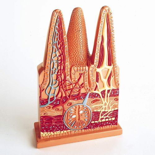
Magnified hundreds of times, three of the more than 5 million villi that line the small intestine are replicated in this vinyl model depicting their essential role in digestion and nutrient absorption.
The Denoyer-Geppert Intestinal Villi model accurately represents three of the five million villi that line the walls of the 27-foot small intestine in humans.
Villi exist to increase the surface area available for food absorption; this area has been estimated at approximately eleven square yards. Also, under some conditions, food may pass through the small intestine rapidly; the villi make the most of the time that the digested material is in contact with the intestine.
The intestinal villi (1) project from the surface of the mucous membrane over the folds and between them. They have cores of lamina propria (8), which is composed of loose connective tissue. Each villus contains a vascular capillary network (3), a central lacteal (7), and nerve fibers.
Lymphatic tissue abounds in the lamina propria and may appear as solitary nodules or in groups of nodules called Peyer’s patches. The flow of lymph is controlled by valves so it can pass out of the intestine only. This lymphatic system makes up one of the two important routes that absorbed particles travel to enter the blood stream.
The second consists of the vascular system; here, one arteriole (5) enters each villus splitting into capillaries and is collected by venules (4).
The crypts of Lieberkühn (6) open between the villi. They produce enzymes, mucus and possibly a hormone. There are goblet cells (21) in the crypts among the columnar epithelium (20).
Related Searches
- Anatomy model
- Demonstration anatomical model
- Teaching anatomy model
- Bone anatomical model
- Flexible anatomical model
- Intracranial anatomical model
- Denture model
- Plastic anatomy model
- Transparent anatomical model
- Oral anatomical model
- Whole body anatomical model
- Vascular model
- Training vascular model
- Leg anatomy model
- Vertebral column model
- Digestive system model
- Pelvic anatomical model
- Cardiac anatomical model
- Nervous system model
- Circulatory system vascular model
*Prices are pre-tax. They exclude delivery charges and customs duties and do not include additional charges for installation or activation options. Prices are indicative only and may vary by country, with changes to the cost of raw materials and exchange rates.


