- Primary care
- General practice
- Intervetebral disc model
- Dynamic Disc Designs Corp.
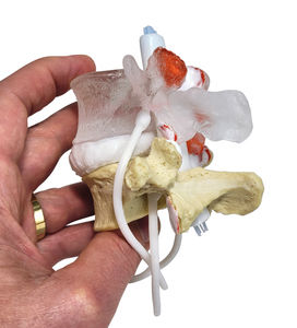
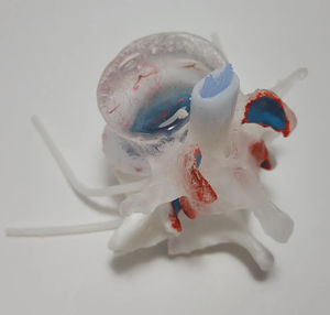
Lumbar vertebra anatomical model Academic LxH intervertebral discfor teachingwith nerves

Add to favorites
Compare this product
Characteristics
- Area of the body
- lumbar vertebra, intervertebral disc
- Procedure
- for teaching
- Accessories
- with nerves
- Color
- white
- Other characteristics
- flexible, transparent
Description
New features include:
circumferential (diffuse) disc bulge
superimposed disc protrusion
limacon shaped annulus
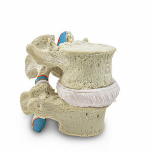
peripherally exposed calcified endplate
elastomeric white articular cartilage
subchondrial bone exposed with hyaline fibrillation
bone coloured L5
white superior endplate matching the colour of the articular cartilage
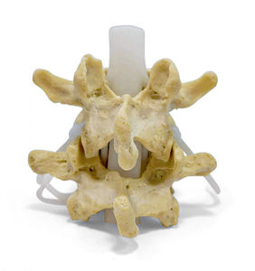
New cauda equina
The anatomical features that remain (from the previous model):
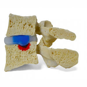
flexible and dynamic herniating (or prolapse) nucleus pulposus. This is achieved through a realistic 2-part intervertebral disc with 6 degrees of freedom. Nuclear migration upon manual compression through a torn annulus fibrosus explaining pain generators under load.
right posterior-lateral radial and circumferential(concentric) fissure
transparent L4
randomly scattered and embedded black nuclear structures to show nuclear shifting dynamics through the L4 view lens easily
L5 superior endplate pores (black)
L5 superior endplate lesion (red)
vasculature in L4 vertebral body (red)
facet subchondrial vascularization (red)
facet tropism (L5 inferior)
VIDEO
Catalogs
Other Dynamic Disc Designs Corp. products
Lumbar Single Level
Related Searches
- Anatomy model
- Demonstration anatomical model
- Teaching anatomy model
- Surgical anatomical model
- Bone anatomical model
- Flexible anatomical model
- Transparent anatomical model
- Vertebral column model
- White anatomical model
- Pelvic anatomical model
- Nervous system model
- Anatomical model with nerves
- Lumbar vertebral column model
- Cervical model
- Nervous anatomy model
- Intervetebral disc model
- Beige anatomical model
- Injection model
- Puncture anatomy model
- Gynecological anatomical model
*Prices are pre-tax. They exclude delivery charges and customs duties and do not include additional charges for installation or activation options. Prices are indicative only and may vary by country, with changes to the cost of raw materials and exchange rates.


