- Products
- Skeleton model
- Erler-Zimmer
Skeleton anatomy model 6059footfor teachingwith musculature
Add to favorites
Compare this product
Characteristics
- Area of the body
- skeleton, foot
- Procedure
- for teaching
- Accessories
- with musculature
Description
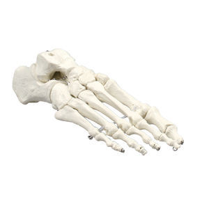
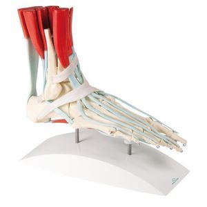
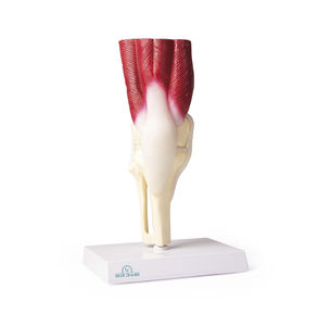
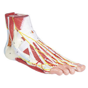
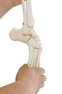
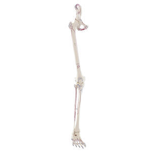
This life-size model of a right human foot shows the anatomy of the musculoskeletal system in detail and is a great aid to understanding and learning the function of muscles and ligaments.
Shown are the major ligaments and muscles:
Extensor hallucis longus and brevis, Extensor digitorum longus and brevis, Fibularis peroneus tertius and brevis, Fibularis peroneus longus and brevis, Abductor hallucis, Digitorum pedis, Flexor digitorum longus and brevis, Tibialis posterior, Flexor digitorum longus, Flexor hallucis longus and Calcaneus muscle. Lumbricales pedis I-IV, Flexor digiti minimi brevis, Abductor digiti minimi
The following retinacula can be seen in the model:
Ret. musculorum extensorum inferius, Ret. musculorum fibularium, Ret. musculorum flexorum.
The muscles and tendons are made of soft material.
Catalogs
No catalogs are available for this product.
See all of Erler-Zimmer‘s catalogs*Prices are pre-tax. They exclude delivery charges and customs duties and do not include additional charges for installation or activation options. Prices are indicative only and may vary by country, with changes to the cost of raw materials and exchange rates.


