- Primary care
- General practice
- Tooth model
- Erler-Zimmer
Skull model 4860jawteethoral
Add to favorites
Compare this product
Characteristics
- Area of the body
- skull, jaw, teeth, oral
- Application
- for dental surgery
- Simulated pathology / condition
- with TMD
Description
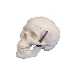
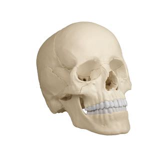
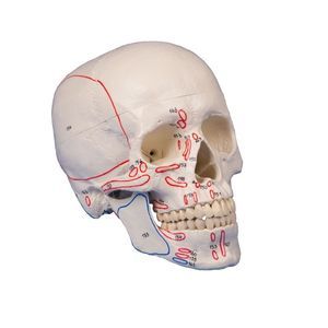
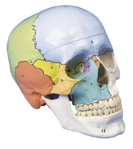
In addition to the detailed reproduction of the skull structures, this first-class model of a human skull focuses specifically on teeth and jaws and is therefore the perfect teaching and demonstration tool for the field of dentistry and oral surgery.
The front half of the upper and lower jaw can be removed to reveal teeth with roots, unilateral nerves and blood vessels as well as the maxillary sinuses. The skull is not only suitable as a perfect aid for learning anatomy, but also as an explanatory aid for oral surgery and implantology.
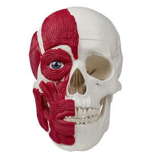
This special design serves to illustrate the TMD syndrome. In temporomandibular dysfunction, there is a dysfunction in the interaction between temporomandibular joints, masticatory muscles and teeth. The wear of the tooth enamel due to grinding, wear of the discus as well as relevant masticatory muscles M. masseter, pars superficialis, pars profundus as well as the relatively newly discovered pars coronoidea are shown. All three muscles are detachable.
Catalogs
No catalogs are available for this product.
See all of Erler-Zimmer‘s catalogsRelated Searches
- Erler-Zimmer anatomical model
- Erler-Zimmer training anatomical model
- Erler-Zimmer teaching anatomical model
- Surgical anatomical model
- Erler-Zimmer bone model
- Erler-Zimmer skull model
- Erler-Zimmer flexible anatomical model
- Denture model
- Plastic anatomy model
- Transparent anatomical model
- Dental anatomical model
- Oral anatomical model
- Erler-Zimmer body anatomical model
- Erler-Zimmer spine anatomical model
- Erler-Zimmer leg anatomical model
- White anatomical model
- Erler-Zimmer heart model
- Erler-Zimmer pelvis model
- Erler-Zimmer digestive system model
- Erler-Zimmer nervous system model
*Prices are pre-tax. They exclude delivery charges and customs duties and do not include additional charges for installation or activation options. Prices are indicative only and may vary by country, with changes to the cost of raw materials and exchange rates.


