- Primary care
- General practice
- Skull model
- Erler-Zimmer
Skull model MP1620for teaching
Add to favorites
Compare this product
Characteristics
- Area of the body
- skull
- Procedure
- for teaching
Description
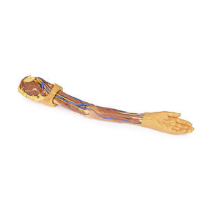
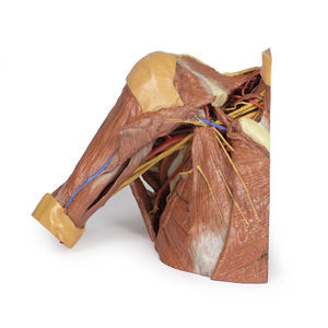
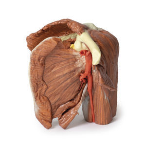
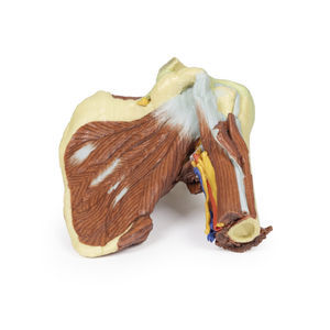
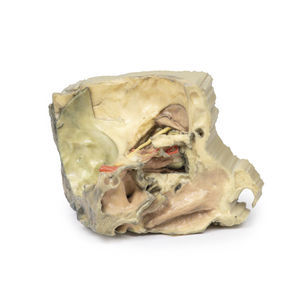
This 3 part 3D printed model derived from CT data highlights the complex anatomy of the temporal bone including bone ossicles, canals, chambers, foramina and air spaces. In addition, the spatial relations between temporal bone and other structures of otological importance, i.e. carotid artery, dural venous sinuses, related nerves and the dura mater are indicated. Internal casts (endocasts) of the bony chambers and canals have been created to aid visualisation of the internal anatomy of the temporal bone.
The model set consists of three parts:
- Part 1 Skull Preparation
- Part 2 The Petrous Part Of The Temporal Bone
- Part 3 The Auditory And Vestibular Apparatus
Detailed anatomical description on request.
Catalogs
Catalog 2021
360 Pages
3D Anatomy Series
9 Pages
Related Searches
- Erler-Zimmer anatomical model
- Erler-Zimmer training anatomical model
- Erler-Zimmer teaching anatomical model
- Surgical anatomical model
- Erler-Zimmer bone model
- Erler-Zimmer skull model
- Erler-Zimmer flexible anatomical model
- Denture model
- Plastic anatomy model
- Transparent anatomical model
- Dental anatomical model
- Oral anatomical model
- Erler-Zimmer body anatomical model
- Erler-Zimmer spine anatomical model
- Erler-Zimmer leg anatomical model
- White anatomical model
- Erler-Zimmer heart model
- Erler-Zimmer pelvis model
- Erler-Zimmer digestive system model
- Erler-Zimmer nervous system model
*Prices are pre-tax. They exclude delivery charges and customs duties and do not include additional charges for installation or activation options. Prices are indicative only and may vary by country, with changes to the cost of raw materials and exchange rates.


