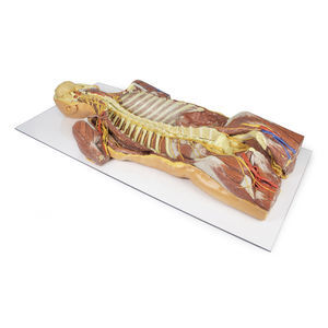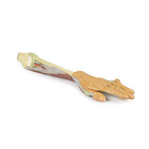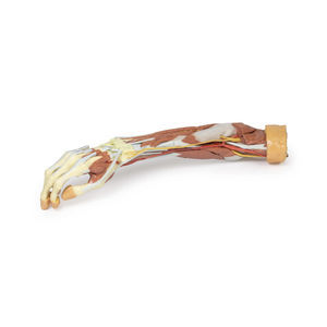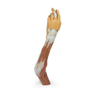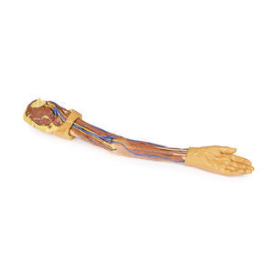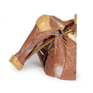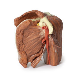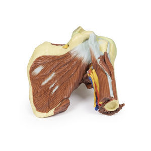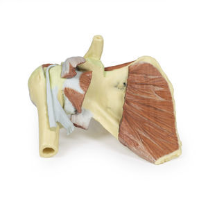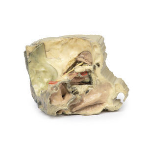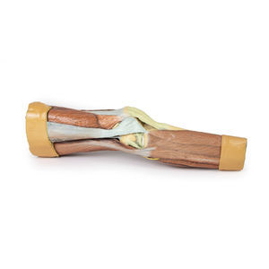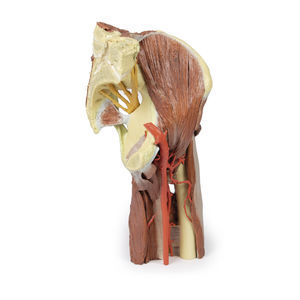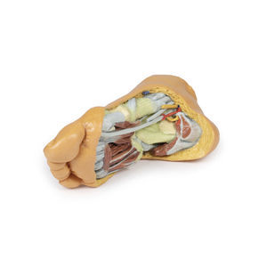
Foot anatomical model MP1900for teaching
Add to favorites
Compare this product
Characteristics
- Area of the body
- foot
- Procedure
- for teaching
Description
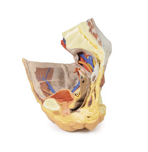
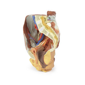
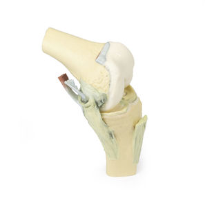
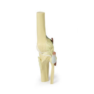
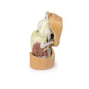
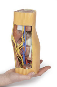
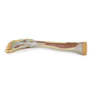
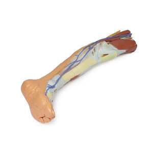
This 3D print records the anatomy of a right distal leg and the deep structures of the plantar surface of the foot. Proximally, the tibia, fibula, interosseous membrane, and leg muscles are discernable in cross-section. Medially, at the level of the ankle joint, the long tendons of the dorsi- and plantar-flexors are visible superficial to the capsular and extra capsular ligaments.
Catalogs
Catalog 2021
360 Pages
3D Anatomy Series
9 Pages
Related Searches
- Erler-Zimmer anatomical model
- Erler-Zimmer training anatomical model
- Erler-Zimmer teaching anatomical model
- Surgical anatomical model
- Erler-Zimmer bone model
- Erler-Zimmer flexible anatomical model
- Erler-Zimmer skull model
- Denture model
- Transparent anatomical model
- Plastic anatomy model
- Dental anatomical model
- Oral anatomical model
- Erler-Zimmer body anatomical model
- Erler-Zimmer spine anatomical model
- Erler-Zimmer leg anatomical model
- Erler-Zimmer heart model
- Erler-Zimmer digestive system model
- Erler-Zimmer pelvis model
- Erler-Zimmer nervous system model
- White anatomical model
*Prices are pre-tax. They exclude delivery charges and customs duties and do not include additional charges for installation or activation options. Prices are indicative only and may vary by country, with changes to the cost of raw materials and exchange rates.


