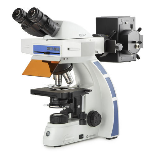
Fluorescence microscope Oxionopticalbiologicalmedical
Add to favorites
Compare this product
Characteristics
- Type
- optical
- Applications
- biological, medical
- Ergonomics
- upright
- Microscope head
- binocular
- Quality of the objectives
- plan, semi-apochromat
- Observation technique
- fluorescence
- Configuration
- benchtop
- Light source
- LED
- Magnification
4 unit, 10 unit, 40 unit, 100 unit
- Resolution
365 nm, 455 nm, 560 nm
Description
For biological and medical applications fluorescence microscopes are used to observe certain parts of living cells and tissues with help of fluorophores. One or more fluophores are added to the parts of the specimen to be observed. When the fluorophores are exposed to so-called exicitation light, the excitation energy is absorbed by the fluorophore that start emitting so-called emission light that can be observed by the microscope. One can distinguish tumor cells from other cells, follow biological processes or prove the presence or absence of antibodies, etc
The Oxion microscopes for fluorescence are equipped with an epi-illumination with a 100 W mercury vapor lamp. The fluorescence attachment have a rail for maximum 4 filter cubes. The FluoLed model is equipped with a 455 nm LED and one filter cube, especially developed for efficient and fast detection of tuberculosis by the Auramine-O fluorochrome. Other FluoLED wavelengths are also available
Highlights
Reversed nosepiece for maximum 5 objectives
Integrated mechanical stage
Height adjustable abbe n.a. 1.25 condenser with iris diaphragm and filter holder
WF 10x/22 mm eyepieces
Plan, plan fluarex and plan semi apochromatic fluarex ioS objectives
Coaxial coarse and fine adjustments
3 W lED transmitted illumination
Fluorescence attachment for maximum 4 filter blocks and with 100 W mercury vapor incident illuminati
Ergonomic stand and head
EYEPIECES
• HWF 10x/22 mm eyepieces
• All eyepieces can be secured by an Allen screw
HEAD
• Binocular and trinocular Siedentopf type heads are with 30° inclined tubes
• Interpupillary distance from 50 to 75 mm
Catalogs
Euromex Oxion
6 Pages
Related Searches
- Microscopy
- Compound microscope
- Laboratory microscope
- Tabletop microscope
- Microscope with LED light
- CMOS camera
- USB camera
- LED illuminator
- Digital microscope
- Biological microscope
- Microscopy camera
- Binocular microscope
- High-definition camera
- Trinocular microscope
- Fluorescence microscope
- Compact illuminator
- High-definition microscope
- Compact microscope
- Medical microscope
- Achromatic microscope
*Prices are pre-tax. They exclude delivery charges and customs duties and do not include additional charges for installation or activation options. Prices are indicative only and may vary by country, with changes to the cost of raw materials and exchange rates.


