- Primary care
- General practice
- Tooth model
- FOSHAN JINGLE MEDICAL EQUIPMENT CO., LTD
- Products
- Catalogs
- News & Trends
- Exhibitions
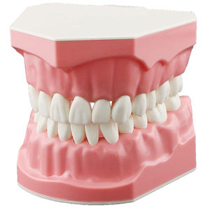
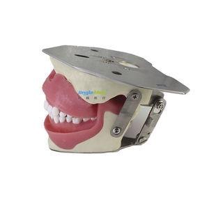
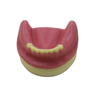
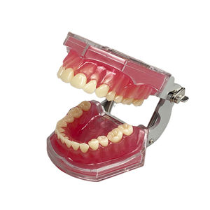
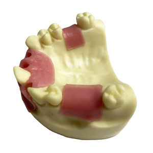
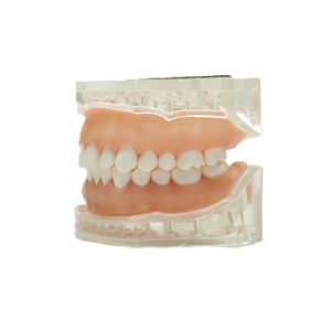
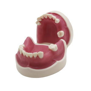
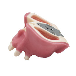
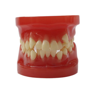
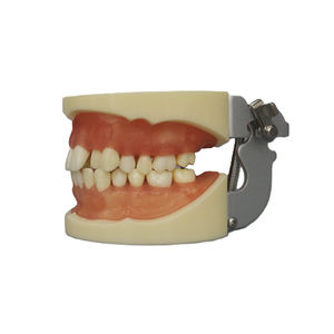
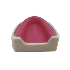
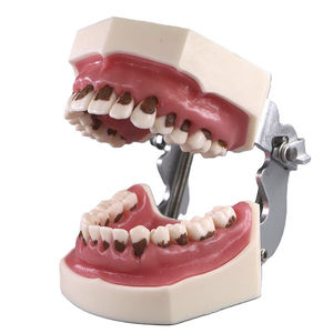
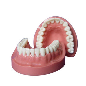
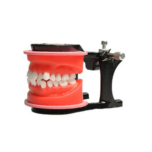
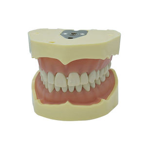
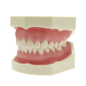
Mouth anatomical model JG-Y29teethdentaldenture

Add to favorites
Compare this product
Characteristics
- Area of the body
- mouth, teeth, denture, dental
- Procedure
- training, for demonstration
- Other characteristics
- interactive
Description
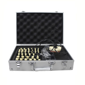
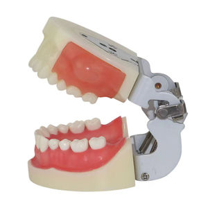
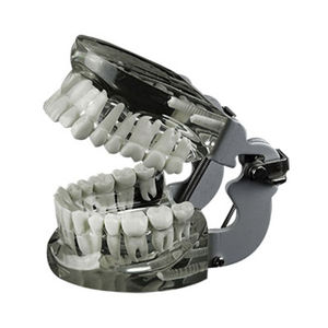
Radiology model developed specifically for for x-ray training. Each tooth is anatomical shaped and permanently fixed-in with full pulp and canal. These teeth are made of radiolucent material with special opaque qualities for in-school or in-office training.
Desciption
Dental Model X-Rays Practice Demonstration Model is a specialized training tool used in dental education to simulate the practice and interpretation of dental X-rays. This model allows students and dental professionals to practice taking and analyzing dental X-rays in a controlled and realistic environment.
Features and Benefits:
X-ray Simulation: The model is designed to simulate the appearance and positioning of teeth and surrounding structures on dental X-rays. It accurately replicates the anatomical features and radiographic characteristics seen in real patient X-rays, providing a lifelike representation for practicing X-ray techniques.
Hands-on Practice: The demonstration model allows students to practice taking dental X-rays using X-ray equipment. They can position the X-ray film or digital sensor correctly in the model's mouth, adjust exposure settings, and capture X-ray images. This hands-on practice helps students develop the necessary skills and proficiency in dental radiography.
Educational Tool: The demonstration model serves as an educational tool to teach and demonstrate dental radiography techniques.Instructors can use the model to explain X-ray positioning, exposure settings, and interpretation criteria. It allows for interactive learning, discussions, and feedback, enhancing students' understanding of dental X-rays.
VIDEO
Catalogs
No catalogs are available for this product.
See all of FOSHAN JINGLE MEDICAL EQUIPMENT CO., LTD‘s catalogsExhibitions
Meet this supplier at the following exhibition(s):
Other FOSHAN JINGLE MEDICAL EQUIPMENT CO., LTD products
Dental Model
Related Searches
- Anatomy model
- Demonstration anatomical model
- Teaching anatomy model
- Surgical anatomical model
- Bone anatomical model
- Intracranial anatomical model
- Flexible anatomical model
- Denture model
- Transparent anatomical model
- Dental anatomical model
- Oral anatomical model
- Implantology anatomical model
- Silicone anatomy model
- Foam anatomical model
- Mandibular model
- Jaw anatomical model
- Gingival anatomical model
- Suture anatomy model
- Maxilla anatomical model
- Resin anatomical model
*Prices are pre-tax. They exclude delivery charges and customs duties and do not include additional charges for installation or activation options. Prices are indicative only and may vary by country, with changes to the cost of raw materials and exchange rates.


