- Secondary care
- Ophthalmology
- Scheimpflug camera
- Heidelberg Engineering
Scheimpflug camera HRT3 RCMtabletop

Add to favorites
Compare this product
Characteristics
- Type of instrument
- Scheimpflug camera
- Ergonomics
- tabletop
Description
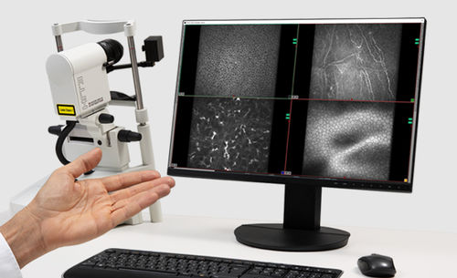
HRT3 RCM is a compact ophthalmic device that uses confocal scanning laser microscopy to provide high-resolution images of the cornea and external ocular structures, such as the conjunctiva or the limbus.
Scanning the cornea with a field of view of up to 400 x 400 μm, HRT3 RCM allows you to acquire unique en face images of corneal cells and structures, identify keratocytes subpopulations, and visualize details of the corneal subbasal nerve plexus.
Navigate through the cornea at the cellular level and select your preferred scanning depth for a comprehensive in vivo assessment of all corneal layers – from epithelium to endothelium.
The comprehensive assessment of the cornea and other external ocular structures with HRT3 RCM can aid you in the diagnosis and monitoring of corneal abnormalities, pre- and post-surgery evaluation, the assessment of dry eye disease, or in the analysis of corneal nerve structure in diabetic patients.
Epithelium – 30 μm
Examine the epithelium in high detail, including the subdifferentiation of epithelial cell layers.
Corneal Subbasal Nerve Plexus – 62 μm
Analyze the fine details of the subbasal nerve plexus to evaluate the corneal nerve structure in diabetic patients.
Stroma – 300 μm
With the high-resolution en face images, you can identify keratocytes subpopulations and precisely analyze corneal treatment outcomes.
Endothelium – 572 μm
The semi-automated endothelial cell count provides additional information about the morphology of this layer. After manually marking the endothelial cells in an appropriate frame, the cell density (cells/mm2) is automatically calculated.
VIDEO
Catalogs
No catalogs are available for this product.
See all of Heidelberg Engineering‘s catalogs*Prices are pre-tax. They exclude delivery charges and customs duties and do not include additional charges for installation or activation options. Prices are indicative only and may vary by country, with changes to the cost of raw materials and exchange rates.


