- Laboratory
- Laboratory medicine
- FE-SEM microscope
- Hitachi High-Tech Europe GmbH
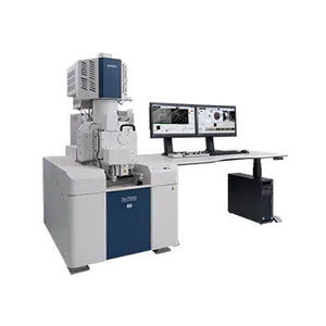
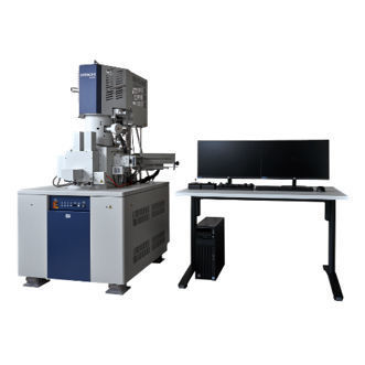
Field emission scanning electron microscope SU8700laboratoryfor researchfloor-standing

Add to favorites
Compare this product
Characteristics
- Type
- field emission scanning electron
- Applications
- laboratory, for research
- Configuration
- floor-standing
- Electron source
- Schottky field emission
- Detector type
- back-scattered electron, secondary electron, energy-dispersive X-ray detector
- Other characteristics
- ultra-high resolution, automated, variable pressure scanning
- Magnification
Min.: 20 unit
Max.: 2,000,000 unit
- Resolution
0.6 nm, 0.9 nm
Description
Equipped with a 150mm sample airlock as standard, the SU8700 offers high sample throughput even for larger samples and a constantly clean sample chamber environment for low-contamination, high-resolution imaging. In addition, the sample chamber can be opened and evacuated again in a matter of minutes to insert accessories. The sample stage can be moved 110mm in X and Y directions. An integrated colour camera enables image-based navigation. There are plenty of connection options for 2 x EDX, EBSD, STEM, inert gas sample transfer, plasma cleaner and other accessories are available.
Product features:
- Durable and stable Hitachi Schottky field emitter with up to 200nA probe current
- Brilliant imaging performance - without the need for a decelerating field on the sample - from 100V (10V option) up to 30kV acceleration voltage. EDX analysis and high-resolution imaging with all detectors are possible at 6mm working distance
- Reliable automatic functions such as adaptation to user defined optical conditions or 2D autofocus and autostigmator enable practical use of the superior equipment capabilities
- A 150mm diameter sample airlock is supplied as standard. It enables fast specimen exchange while keeping the chamber vacuum clean
VIDEO
Catalogs
Exhibitions
Meet this supplier at the following exhibition(s):


Other Hitachi High-Tech Europe GmbH products
Electron Microscopes (SEM/TEM/STEM)
Related Searches
- Hitachi microscope
- Hitachi laboratory microscope
- Hitachi benchtop microscope
- Biological microscope
- Hitachi research microscope
- Hitachi high-resolution microscope
- Compact microscope
- Medical microscope
- Inspection microscope
- Dark field microscope
- 3D microscope
- Multipurpose microscope
- Hitachi floor-standing microscope
- Hitachi SEM microscope
- Materials research microscope
- Super-resolution microscope
- TEM microscope
- STEM microscope
- Hitachi automated microscope
- Hitachi FE-SEM microscope
*Prices are pre-tax. They exclude delivery charges and customs duties and do not include additional charges for installation or activation options. Prices are indicative only and may vary by country, with changes to the cost of raw materials and exchange rates.


