- Primary care
- General practice
- Heart model
- JMC Corporation

- Company
- Products
- Catalogs
- News & Trends
- Exhibitions
Heart model trainingbiopsytransparent

Add to favorites
Compare this product
Characteristics
- Area of the body
- heart
- Procedure
- training, biopsy
- Other characteristics
- transparent
Description
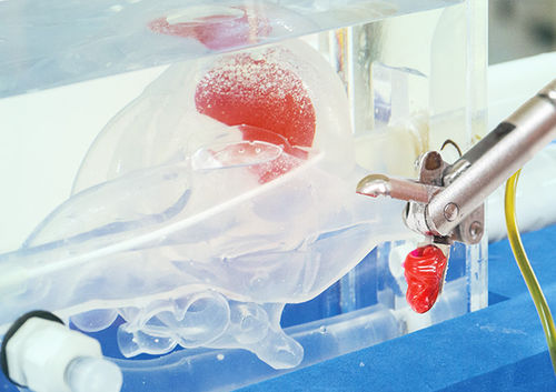
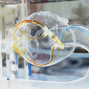
With this model, the myocardial biopsy procedure can be simulated under X-ray fluoroscopy, similar to the set-up in a real cath lab. The transparent heart model enables one to practice the procedure by confirming the direction of the sheath and forceps through both an X-ray image and a camera image.
Using the X-ray image, it is possible to determine if the forceps are facing towards the free wall. The compact camera with a flexible arm can provide a clear image from various angles.
As the material used to simulate the ventricular septum is different from that of the ventricular free wall, it is easy to confirm whether the tissue was removed from the appropriate area after the procedure.
VIDEO
Catalogs
No catalogs are available for this product.
See all of JMC Corporation‘s catalogsRelated Searches
- Anatomy model
- Demonstration anatomical model
- Surgical anatomical model
- Transparent anatomical model
- Vascular model
- Training vascular model
- Cardiac anatomical model
- Transparent vascular model
- Circulatory system vascular model
- Blood vessels vascular model
- Circulatory system model
- Artery vascular model
- Vascular surgery vascular model
- Cardiac vascular model
- Catheterization vascular model
- Coronary artery anatomical model
- Coronary arteries vascular model
- Vascular surgery model
- Aortic valve model
- Cardiovascular catheterization vascular model
*Prices are pre-tax. They exclude delivery charges and customs duties and do not include additional charges for installation or activation options. Prices are indicative only and may vary by country, with changes to the cost of raw materials and exchange rates.


