- Secondary care
- Ophthalmology
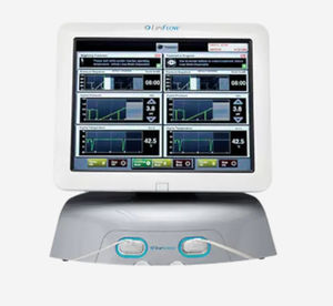
- Meibography dry eye diagnosis system
- Johnson & Johnson Vision
Meibography dry eye diagnosis system TearScience™ LipiScan™
Add to favorites
Compare this product
Characteristics
- Method
- meibography
Description
With a small footprint and user-friendly design, the TearScience LipiScan with Dynamic Meibomian Imaging (DMI) was designed to make high-definition meibography accessible for any practice.
Key Features
High-definition meibomian imaging
Provides enhanced view of meibomian gland structure
Versatile design
Small and light weight
Maximize workflow efficiency
Easy facility integration with meibomian gland imaging in about 10 seconds
How TearScience™ LipiScan™ works
Workflow maximization with fast capture of meibomian gland images
Small footprint and lightweight (25 lbs) for optimal versatility
Fast and intuitive operation for seamless integration into routine workups
Renders high-definition image of meibomian gland structure
DICOM compatibility to export images to EMR
Dynamic meibomian imaging
Images for illustrative purposes only. Actual results may vary.
Dynamic Illumination
Surface lighting originates from multiple light sources to minimize reflection.
Adaptive Transillumination
Changes to the light intensity across the surface of the illuminator compensate for the lid thickness variations between patients.
Dual-Mode DMI
Dynamic Illumination offers an enhanced view of meibomian gland structure
Catalogs
No catalogs are available for this product.
See all of Johnson & Johnson Vision‘s catalogsRelated Searches
- Diode laser
- Fixed ophthalmic examination
- Tabletop ophthalmic examination instrument
- Intraocular lens
- Ophthalmic laser
- Refractometer ophthalmic examination
- Automatic refractometer
- Monofocal intraocular lens
- Trifocal intraocular lens
- Capsulotomy laser
- Dry eye diagnosis system
- Phacoemulsifier
- Meibography dry eye diagnosis system
- Floor-standing laser
- Cataract surgery laser
- Femtosecond laser
- Vitrectome
- Excimer laser
- Interferometry dry eye diagnosis system
*Prices are pre-tax. They exclude delivery charges and customs duties and do not include additional charges for installation or activation options. Prices are indicative only and may vary by country, with changes to the cost of raw materials and exchange rates.


