- Secondary care
- Ophthalmology
- Specular microscope
- Konan Medical USA

- Products
- Catalogs
- News & Trends
- Exhibitions
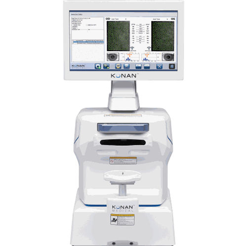
Pachymeter CellChek® XLspecular microscopespecular microscopic pachymetrytabletop

Add to favorites
Compare this product
Characteristics
- Type of instrument
- pachymeter, specular microscope
- Examination
- specular microscopic pachymetry
- Ergonomics
- tabletop
Description
Endothelial cell morphology is like a ‘canary in the coal mine’…a potential warning that the structural stability of the endothelium has been affected, possibly by surgical procedures, disease, trauma or contact lenses.
Clinical Applications
Glaucoma, cataract, & refractive surgery.
Corneal disease management.
Contact/specialty contact lens fittings.
Routine eye care
Clinical Benefits
Visualize endothelial cells with 40x magnification compared to slit lamp bio-microscopy
Identify pre-existing low density and dystrophies that may affect positive surgical outcomes
Confirm recommended cell density/morphology for scleral/specialty contact lenses
CellChek SL | XL Models
Gently used devices come with 'same-as-new' warranty. Examples include trade show floor / demo units, clinical trial returns and trade-ins. Limited availability. Only available in the USA.
Clinical Benefits
Cataract Surgery and Premium IOLs
Low endothelial cell counts and pre-existing dystrophies can markedly reduce the potential for positive surgical outcomes from an otherwise uneventful cataract surgery. Surgeons are finding these data points critical when recommending premium IOLs: verify and document pre-operatively that the cornea is not suspect to more likely post-op complications difficult to explain with the investment in premium IOLs.
General Corneal Health Assessment
Konan specular microscopy is an invaluable tool to screen for corneal diseases as such as Fuchs’ Dystrophy, keratoconus, other corneal dystrophies, and trauma. You won’t believe what you’ve been missing.
VIDEO
*Prices are pre-tax. They exclude delivery charges and customs duties and do not include additional charges for installation or activation options. Prices are indicative only and may vary by country, with changes to the cost of raw materials and exchange rates.


