
- Company
- Products
- Catalogs
- News & Trends
- Exhibitions
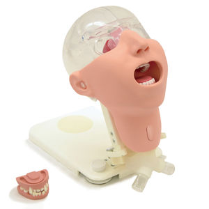
Otoscopy patient simulator M82Ahead

Add to favorites
Compare this product
Characteristics
- Application
- for otoscopy
- Form
- head
Description
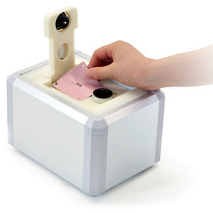
Practical fundus examination simulator with 10 clinical images and variations
Features -
1. 10 fundus slide cases of common eye diseases
2. Real clinical images
3. Lenses are used for a part of the eyeball, and reproduces the visual axis close to human.
4. You can examine the optic fundus with any ophthalmoscope available on the market.
Provides you with the simulation of an actual examination.
5. It is possible to change the degree of dilation and contraction of the pupil in 3 steps (2, 3.5,5mm) offering different degrees of challenges. (*M82A is 2 steps; 3.5, 8mm)
6. The soft and supple material allows simulations of a real examination, in ways such as pulling up the eyelid.
Training skills / Applications - Funds examination with direct ophthalmoscope
Case / Pathology - Normal fundus / Hypertensive retinopathy:-Grade 3 arteriolar vasoconstriction -Grade 1 arteriolosclerosis / -hemorrhages and cotton wool spots / -simple vein concealment / Diabetic retinopathy:microaneurysm,hemorrhages and hard exudates / Papilloedema(chronic phase)/ Papilloedema(acute phase)/ Glaucomatous optic atrophy:glaucomatous optic disc cuppping and nerve fiber defect / Rerinal vein occlusion(acute phase):flame-shaped hemorrhage and cotton wool spots / Rerinal vein occlusion(post rerinal laser photocoagulation)/ Toxoplasmosis:retinochoroiditis / Age-related macular degeneration:macular exudates and subretinal hemorrhage / ※M82A includes two normal fundus slides.
Size (approx.) - W42x D22×H39㎝ / W16.5×D8.7×H15.4in
Packing size (approx.) - M82:W49x D29x H53㎝/W19x D11x H20in
M82A:W48xD27x36cm/W19xD11xH14in
Weight (approx.) - 2㎏/4.4lbs
Packing weight (approx.) - 4kg/8lb
VIDEO
Catalogs
Related Searches
- Demonstration simulator
- General care medical simulator
- Training manikin
- Upper body simulator
- Surgical simulator
- Patient simulation unit
- Pad simulator
- Injection simulator
- Portable simulation trainer
- Lower body simulator
- General care training manikin
- Vital sign simulator
- Upper body training manikin
- Emergency care simulation unit
- Woman simulator
- Puncture simulator
- Obstetrical/gynecological simulator
- Suture simulator
- Plastic simulator
- Anatomy simulation trainer
*Prices are pre-tax. They exclude delivery charges and customs duties and do not include additional charges for installation or activation options. Prices are indicative only and may vary by country, with changes to the cost of raw materials and exchange rates.


