- Laboratory
- Sample management
- Vibrating blade microtome
- Leica Biosystems
- Company
- Products
- Catalogs
- News & Trends
- Exhibitions
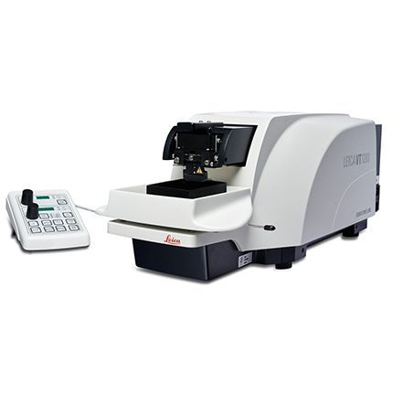
Vibrating blade microtome Leica VT1200semi-automaticfor research

Add to favorites
Compare this product
Characteristics
- Type
- vibrating blade
- Operation
- semi-automatic
- Applications
- for research
Description
The semi-automatic Leica VT1200 vibrating blade microtome is designed for sectioning fresh specimens in neuropathology (fresh brain slicing), neurophysiology (patch-clamping), and electrophysiology.
The instrument is recommended for users who prefer to control sectioning parameters such as section thickness and cutting stroke manually for each individual section. To achieve sections that retain viable cells on the section surface, the vertical deflection of the blade can be measured with the Leica Vibrocheck measurement device and minimized with the innovative blade holder.
The instrument was designed in collaboration with Prof. Dr. Peter Jonas (previously at the Physiology department of the University of Freiburg, Germany, now at Institute of Science and Technology, Klosterneuburg, Austria) and his former group.
Manually control sectioning
Designed for users who prefer to manually control sectioning parameters such as section thickness and cutting stroke for each individual section.
Minimal vertical deflection
Minimal vertical deflection minimizes specimen damage during sectioning and results in more viable cells on the section surface.
Magnetic specimen holders
Magnetic specimen holders support easy specimen handling and reproducible orientation.
VIDEO
Catalogs
Vibrating Blade Microtomes
8 Pages
Leica VibratomeTM Series
8 Pages
Related Searches
- Leica sample preparation system
- Leica automatic sample preparation system
- Leica laboratory sample preparation system
- Sample box
- Laboratory bath
- Leica benchtop sample preparation system
- Benchtop laboratory bath
- Benchtop water bath
- Thermal printer
- Storage sample box
- Leica tissue processor
- Laboratory printer
- Leica histology sample preparation system
- Leica slide stainer
- Leica biopsy cassette
- Leica microtome
- Sample processor with touchscreen
- Laboratory sample box
- Compact printer
- Modular sample processor
*Prices are pre-tax. They exclude delivery charges and customs duties and do not include additional charges for installation or activation options. Prices are indicative only and may vary by country, with changes to the cost of raw materials and exchange rates.


