- Laboratory
- Laboratory medicine
- Operating microscope video camera
- Leica Microsystems
- Company
- Products
- Catalogs
- News & Trends
- Exhibitions
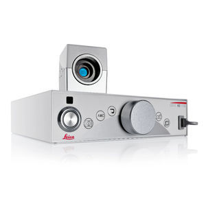
Operating microscope video camera FL800surgicaldigitalinfrared

Add to favorites
Compare this product
Characteristics
- Applications
- surgical, for operating microscopes
- Technology
- digital
- Light source
- fluorescence, infrared, white light
Description
A ruptured cerebral aneurysm is the third most common cause of death in industrial countries.
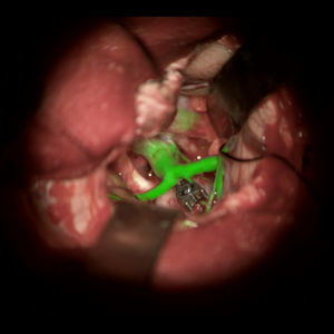
With the Leica FL800, for intra-operative fluorescence-aided videoangiography, surgeons can now determine the patency of vessels.
During surgery, the patient is injected intravenously with ICG (indocyanine green) dye, which is well tolerated and spreads quickly. A near infrared camera then shows the bloodstream in black-and-white directly through the surgical microscope eyepieces and/or on a video monitor.
KEY FEATURES
See blood flow intra-operatively
The Leica FL800 fluorescence module enables the surgeon to observe blood flow through the microscope eyepieces or on the video monitor in real time, without additional measuring apparatus. It is a fast, easy method of checking during surgery whether the aneurysm has been perfectly clipped and whether blood flow is properly flowing through the bypass.
Simple and fast
To change from white light to NIR mode, the surgeon simply pushes a button found on the hand grip of the surgical microscope. Operating the Leica FL800 thus perfectly blends into the surgical workflow. It is simple and fast, this being a main criteria in cerebral aneurysm surgery
Flexible Viewing
Using the Leica FL800 in combination with a digital imaging color module (Leica DI C500 or Leica DI C700) enables the surgeon to display the ICG fluorescence image into the eyepiece. The surgeon can choose how to view the collected data.
VIDEO
Catalogs
Leica M530 OHX
16 Pages
PROvido
8 Pages
Related Searches
- Leica analysis software
- Leica microscope
- Leica optical microscope
- Leica laboratory microscope
- Leica benchtop microscope
- Leica LED microscope
- Leica visualization software
- Control software
- Leica laboratory software
- Windows software
- Leica CMOS camera
- Leica camera with USB port
- Automated software
- Leica LED light source
- Leica digital microscope
- Acquisition software
- Scan software
- Zoom microscope
- Measurement software
- Leica biology microscope
*Prices are pre-tax. They exclude delivery charges and customs duties and do not include additional charges for installation or activation options. Prices are indicative only and may vary by country, with changes to the cost of raw materials and exchange rates.


