- Laboratory
- Laboratory medicine
- Operating microscope video camera
- Leica Microsystems
- Company
- Products
- Catalogs
- News & Trends
- Exhibitions
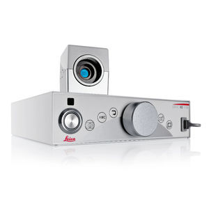
Operating microscope video camera FL400digitalHDfluorescent

Add to favorites
Compare this product
Characteristics
- Applications
- for operating microscopes
- Technology
- digital
- Resolution level
- HD
- Light source
- fluorescent, white light
Description
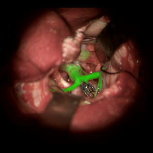
FL400 fluorescence with 5-ALA allows differentiation of tumor tissue from healthy brain tissue to support precise resection.
When resecting a malignant glioma, maximum removal of tumor cells with minimal impact to brain tissue is key to an optimal patient outcome. This is where FL400 fluorescence can provide support. In combination with the active substance 5 aminolevulinic acid (5-ALA), it helps distinguish both the bulk and margins of the tumor more easily. More visual information helps you to confidently perform a resection.
If it glows red, it's tumor
Under FL400 blue light mode, you and your assistant have a bright, high-contrast view of glioma cells. This aids with clear delineation of tumor margins for confident resection in real time.
How does it work?
Before surgery, the patient drinks a 5 aminolevulinic acid (5-ALA) solution. The 5-ALA is selectively taken up by glioma cells and converted into the fluorescent PPIX.
During surgery, you simply activate the FL400 module with a click of a button.
The FL400 module delivers intense, homogeneous blue excitation light, with a well-adjusted observation spectrum.
The PPIX in the tumor glows an intense pinkish red, enabling clear differentiation of tumor cells from healthy tissue
Easy handling for interruption-free work
Quickly and easily switch from white light to blue light fluorescence mode and back, or between fluorescence modes, for an interruption-free workflow.
As the FL400 module is fully integrated into your microscope, changing observation modes requires only the click of a button on the handgrip or footswitch.
VIDEO
Catalogs
Leica M530 OHX
16 Pages
ARveo 8
16 Pages
Related Searches
- Leica analysis software
- Leica microscope
- Leica optical microscope
- Leica laboratory microscope
- Leica benchtop microscope
- Leica LED microscope
- Leica visualization software
- Control software
- Leica laboratory software
- Windows software
- Leica CMOS camera
- Leica camera with USB port
- Automated software
- Leica LED light source
- Leica digital microscope
- Acquisition software
- Scan software
- Zoom microscope
- Measurement software
- Leica biology microscope
*Prices are pre-tax. They exclude delivery charges and customs duties and do not include additional charges for installation or activation options. Prices are indicative only and may vary by country, with changes to the cost of raw materials and exchange rates.


