- Laboratory
- Laboratory medicine
- Optical microscope module
- Leica Microsystems
- Company
- Products
- Catalogs
- News & Trends
- Exhibitions
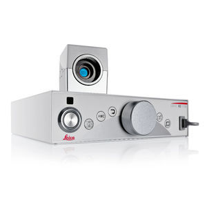
Fluorescence microscope module FL560opticalinfraredmedical
Add to favorites
Compare this product
Characteristics
- Type
- optical, infrared
- Applications
- medical
- Observation technique
- fluorescence, spectral
- Light source
- white light
Description
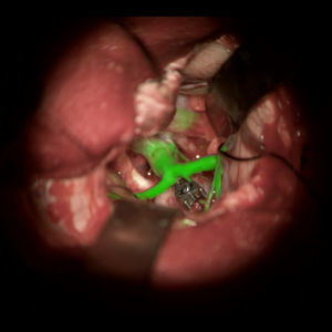
Visualizing certain anatomical and physiological features during neurosurgery can be challenging under white light or near infrared ICG fluorescence.
The FL560 fluorescence module enables simultaneous, real-time observation of both non-fluorescent tissue and fluorescent areas, with clear differentiation and contrast.
One simultaneous view
The proprietary FL560 filter from Leica Microsystems was designed to effectively separate fluorescence excitation light and the observation spectrum. When combined with premium Leica microscope optics, the result is a single real-time view of anatomy and fluorophores with clear differentiation and high contrast.
Which fluorophores can be observed?
Fluorophores with an excitation peak between ~460 nm and ~500 nm (blue)
Fluorescence emission observation comprising the green, yellow and red spectrum in a spectral band above ~510 nm.
Simplify your workflow
Simultaneous anatomical and real-time fluorescence visualization means no need to interrupt workflow to switch back and forth between views.
Thanks to full microscope integration, activating FL560 mode requires just one click of the handgrip or footswitch. If you choose to integrate other fluorescence modes, you can also switch between them with a single click.
Recording & display made easy
Share the view with your team and easily capture brilliant FL560 fluorescence videos in high definition.
HD display of the microscope monitor enables viewing by the whole team in the operating room
HD recording technology in both in white light and FL560 mode is ideal for documentation, presentation and teaching of complex cases
Catalogs
Related Searches
- Leica analysis software
- Leica microscope
- Leica optical microscope
- Leica laboratory microscope
- Leica benchtop microscope
- Leica LED microscope
- Leica visualization software
- Control software
- Leica laboratory software
- Windows software
- Leica CMOS camera
- Leica camera with USB port
- Automated software
- Leica LED light source
- Leica digital microscope
- Acquisition software
- Scan software
- Zoom microscope
- Measurement software
- Leica biology microscope
*Prices are pre-tax. They exclude delivery charges and customs duties and do not include additional charges for installation or activation options. Prices are indicative only and may vary by country, with changes to the cost of raw materials and exchange rates.


