- Laboratory
- Laboratory medicine
- Digital microscope module
- Leica Microsystems
- Company
- Products
- Catalogs
- News & Trends
- Exhibitions
Digital microscope module EnFocusmedicaluprightOCT

Add to favorites
Compare this product
Characteristics
- Type
- digital
- Applications
- medical
- Ergonomics
- upright
- Observation technique
- OCT
- Configuration
- benchtop
Description
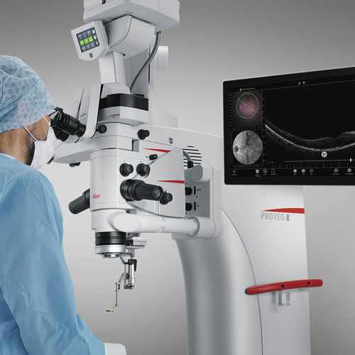
Apply your skills with even greater confidence during complex anterior and posterior segment surgeries with a Leica OCT microscope solution.
EnFocus intraoperative optical coherence tomography (OCT) allows you to see what lies underneath the surface. It gives you the additional real-time information you need for a deeper understanding of how subsurface tissue reacts to your surgical maneuvers.
Our intraoperative OCT imaging system is available fully built into the Proveo 8 ophthalmic microscope. You can enhance the microscope view with OCT imaging at any step during surgery with just a few taps. The Proveo 8 microscope with integrated EnFocus OCT supports you to focus on achieving an optimal patient outcome by providing:
Greater insight into hidden subsurface details
Immediate confirmation on how tissue reacts to your surgical maneuvers
Maximum freedom in viewing and reviewing optimized OCT images
Greater Insight: See more details below the surface
Supplement your microscope view with bright, sharp imaging of previously hidden subsurface tissue details. Intraoperative integration of OCT imaging provides you with additional information so you can get greater insights into ocular pathology during surgery.
Clearly differentiate between artifacts and tissue due to the unique spectrometer technology with dispersion compensation software and a highly sensitive detector that captures more signal
See fine details even through blood in trauma cases thanks to an axial resolution of 2.4 μm in tissue
VIDEO
Related Searches
- Leica analysis software
- Leica microscope
- Leica optical microscope
- Leica laboratory microscope
- Leica benchtop microscope
- Leica LED microscope
- Leica visualization software
- Control software
- Leica laboratory software
- Windows software
- Leica CMOS camera
- Leica camera with USB port
- Automated software
- Leica LED light source
- Leica digital microscope
- Acquisition software
- Scan software
- Zoom microscope
- Measurement software
- Leica biology microscope
*Prices are pre-tax. They exclude delivery charges and customs duties and do not include additional charges for installation or activation options. Prices are indicative only and may vary by country, with changes to the cost of raw materials and exchange rates.


