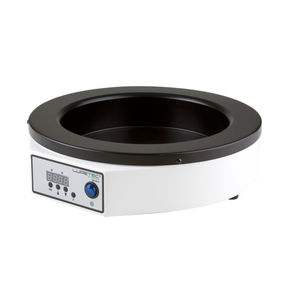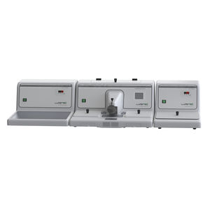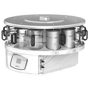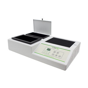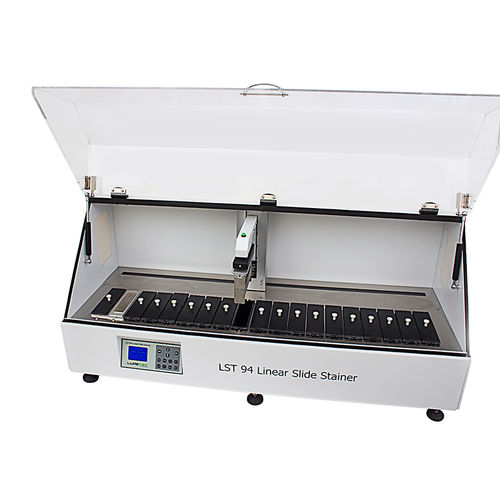
Laboratory sample preparation system LST94for slide stainingtissuelinear array
Add to favorites
Compare this product
Characteristics
- Applications
- laboratory, for slide staining
- Sample type
- tissue
- Configuration
- linear array
- Preparation format
- for microscope slides
Description
Staining of tissuesThe use of dyes is key to view the tissue under the light microscope. After the microtome, the cells and extracellular material are typically transparent and dyes enhance visualization of tissue structures.The dyes applied to stain tissues that were previously set are called non vital stains, such as hematoxylin, eosin, fuchsin, among others.We can also stain cells in culture or cell bodies are still alive; in this case, it is necessary to use dyes called vital that do not cause damage to cells and also does not interfere with cell metabolism. Among them are: the trypan blue, green Janus B, trypan red, methylene blue, neutral red, among others.The automation of this process can be done with Slide Stainers. The model tissue Processor is an apparatus used forroutine staining of animal, plant and human bodytissue. It can be widely used in such institutions ashospitals, scientific research institutes, universities andjudicial departments for clinical pathologic analysis andresearch on animal and plant cells.
Catalogs
No catalogs are available for this product.
See all of LUPETEC‘s catalogsRelated Searches
- Sample processor
- Automated sample preparation system
- Laboratory sample processor
- Laboratory bath
- Benchtop sample processor
- Benchtop laboratory bath
- Benchtop water bath
- Tissue sample processor
- Thermostatic laboratory bath
- Thermostatic water bath
- Histology sample processor
- Slide staining sample processor
- Microtome
- Sample processor with touchscreen
- Modular sample processor
- Microscope slide sample processor
- Rotary microtome
- Digital hotplate
- Embedding sample processor
- Microtome cryostat
*Prices are pre-tax. They exclude delivery charges and customs duties and do not include additional charges for installation or activation options. Prices are indicative only and may vary by country, with changes to the cost of raw materials and exchange rates.


