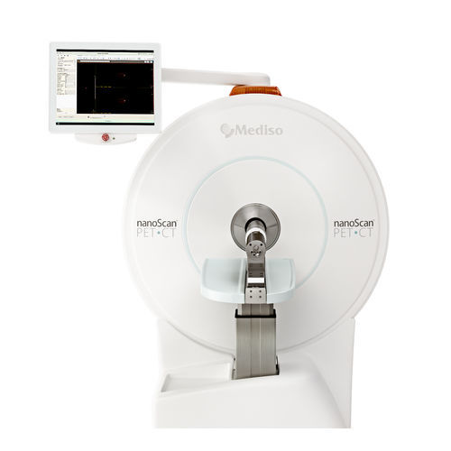
PET preclinical imaging system nanoScan®micro X-ray CTfor small animalsreal-time
Add to favorites
Compare this product
Characteristics
- System type
- PET, micro X-ray CT
- Application
- for small animals
- Other characteristics
- real-time
Description
The nanoScan® PET system is equipped with ultra-fast electronics, and the finest detector crystal pixels in thick layers, ensuring high spatial resolution (down to 700 μm in vivo), and quantitative results, delivered over the widest dynamic range of radioactivity levels from 60 Bq to 80 MBq (1.6 nCi – 2.16 mCi). Combining this with the large axial and transaxial FOV of the PET ring, imaging of large or multiple animals simultaneously is possible, even with 11C- and 15O-labelled radiotracers.
Large ring diameter allows imaging of multiple mice and large animals, while the unique, Tera-Tomo™ 3D iterative reconstruction based on real-time Monte Carlo simulation ensures homogeneous image quality over the entire field of view.
Open access to the animal during PET scanning gives full control over the animal studies.
Features & Benefits
Resolving precise details with 700 μm spatial resolution
Finest (1.12mm×1.12mm) lutetium oxyorthosilicate (LSO) crystal needles provide precise signal localization preserving spatial information in raw data
Tera-Tomo™ 3D PET iterative reconstruction with real-time Monte Carlo based physical modelling unveiling the tiniest details on the image
Large ring diameter and statistical depth of interaction compensation offers homogeneous image quality over the entire field of view
Uncompromised applications with very low level of radioactivity
Thick LSO crystals for excellent sensitivity
Short (3 ns) coincidence time window necessary for improved signal to noise ratio
Advanced corrections (random, scatter, LSO background etc.) ensuring quantification at low activity levels
Catalogs
Related Searches
- Preclinical imaging system
- Medical research preclinical imaging system
- X-rays preclinical imaging system
- Small animal preclinical imaging system
- Real-time preclinical imaging system
- PET preclinical imaging system
- MRI preclinical imaging system
- Cryogen-free preclinical imaging system
- SPECT preclinical imaging system
*Prices are pre-tax. They exclude delivery charges and customs duties and do not include additional charges for installation or activation options. Prices are indicative only and may vary by country, with changes to the cost of raw materials and exchange rates.


