- Company
- Products
- Catalogs
- News & Trends
- Exhibitions
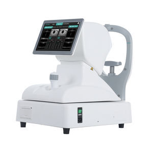
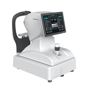
Meibography dry eye diagnosis system DEAinterferometrytear meniscometry

Add to favorites
Compare this product
Characteristics
- Method
- meibography, tear meniscometry, interferometry
Description
Comprehensive 10-in-1 exam capability for ocular surface evaluation
8 mp high resolution camera and outstanding optical design bring high level of detail and clarity
Effortless precision with automated functions: auto NIBUT, tear meniscus height, meibomian gland analysis, and redness detection
Intuitive software that guides you with ease and allows for personalized exam protocols, tailored to your needs
Compact design for easy installation on any slit lamp or handheld use
Auto meibography
Evaluate the meibomian glands with red light, the software provides automatic evaluation of loss area.
Non-invasive breakup time
Automatically analyze break-up area, first and average break-up time for tear stability evaluation.
Interferometry
Record a video of blinking process to observe the surface reflection pattern and dynamics of the tear film.
Tear meniscus height
Automatically evaluate tear meniscus height that is observed on the eyelid margins. Up to 5 measurement points can be taken.
Fluorescein Staining
Evaluate the areas of damage on the ocular surface after application of the fluorescein dye. Compare your images with grading scales incorporated in the software.
Auto Redness
Eye redness could be one of the symptoms of dry eye disease. Automatically compare your images with grading scales incorporated in the software.
Eyelid margin imaging
MGD can cause the glands to become blocked, impacted, and infected. Capture high resolution under white LED illumination, and compare your images with grading scales included in the software .
VIDEO
Catalogs
DEA
8 Pages
Related Searches
- Fixed ophthalmic examination
- Ophthalmic biomicroscope
- Hand-held ophthalmic examination instrument
- Table ophthalmic biomicroscope
- Ophthalmoscope
- Refractometer ophthalmic examination
- Automatic refractometer
- Ophthalmic instrument table
- Electric ophthalmic instrument table
- Height-adjustable ophthalmic instrument table
- Ophthalmic instrument table on casters
- Ophthalmic biometer
- OCT ophthalmoscope
- Retinal imaging instrument
- Dry eye diagnosis system
- Ophthalmic surgery microscope
- Optical ophthalmic biometer
- Meibography dry eye diagnosis system
- Tear meniscometry dry eye diagnosis system
- Interferometry dry eye diagnosis system
*Prices are pre-tax. They exclude delivery charges and customs duties and do not include additional charges for installation or activation options. Prices are indicative only and may vary by country, with changes to the cost of raw materials and exchange rates.


