- Company
- Products
- Catalogs
- News & Trends
- Exhibitions
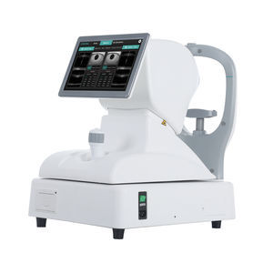
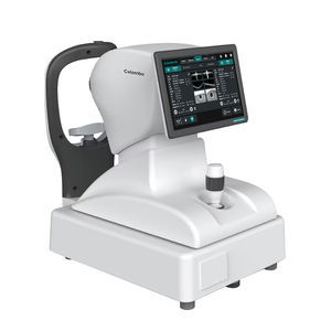
OCT ophthalmoscope Mocean 3000retinal imagingtabletop

Add to favorites
Compare this product
Characteristics
- Type of instrument
- OCT ophthalmoscope
- Examination
- retinal imaging
- Ergonomics
- tabletop
Description
Up to 10x10mm 3D PanoScan
3.1mm scan depth shows better details of the vitreous, retina and choroid
Real-time 45° SLO retinal imaging with eye tracker
AI retina analysis
16mm angle-to-angle scan
Comprehensive analysis of retina, glaucoma and cornea
Real-time SLO with eye tracking
Mocean® 3000 simultaneously acquires OCT images and 45 degrees fundus images based on Scanning Laser Ophthalmoscope (SLO), providing a real-time overview of the retina that allows easy localization of the lesion area before acquisition.
To minimize the artifacts caused by eye drift and micro saccades, Mocean® 3000 uses SLO-based eye tracker, which gives you more confidence in practice.
16mm angle-to-angle analysis
16mm angle-to-angle anterior scan with data analysis.
Deep Choroidal Imaging (DCI) mode
Using Deep Choroidal Imaging for detection of choroidal neovascularization.
Comprehensive software analysis
The Mocean® 3000 system provides 9 scan patterns to help you improve diagnostic efficiency:
Retina (HD line, Six-Radial lines, Multi, 3D Cube 10x10mm)
Glaucoma (Glaucoma Disc for ONH analysis, Glaucoma Macular for GCC analysis)
Cornea (HD line, Six-Radial lines, Angle-to-Angle)
The software analysis features are always up-to-date and free for upgrade (excluding OCTA module).
Methodology Spectral domain OCT
Optical source Super luminescent diode (SLD), 840 nm
Scan speed 50,000 A-scans/s
Axial resolution (optical) 5 microns (optical), 2.7 microns (digital)
Transverse resolution 15 microns (optical), 3 microns (digital)
A-scan depth 3.1 mm
Diopter range - 20 to + 20 diopters
Working distance 30 mm
VIDEO
Catalogs
No catalogs are available for this product.
See all of Moptim‘s catalogsRelated Searches
- Fixed ophthalmic examination
- Tabletop ophthalmic examination instrument
- Ophthalmic biomicroscope
- Hand-held ophthalmic examination instrument
- Table ophthalmic biomicroscope
- Ophthalmoscope
- Refractometer ophthalmic examination
- Automatic refractometer
- Ophthalmic instrument table
- Electric ophthalmic instrument table
- Height-adjustable ophthalmic instrument table
- Ophthalmic instrument table on casters
- Ophthalmic biometer
- OCT ophthalmoscope
- Ophthalmic surgery microscope
- Dry eye diagnosis system
- Retinal imaging instrument
- Optical ophthalmic biometer
- Meibography dry eye diagnosis system
- Tear meniscometry dry eye diagnosis system
*Prices are pre-tax. They exclude delivery charges and customs duties and do not include additional charges for installation or activation options. Prices are indicative only and may vary by country, with changes to the cost of raw materials and exchange rates.


