- Laboratory
- Laboratory medicine
- Staining solution reagent
- Nanjing Liming Bio-products Co., Ltd.

- Company
- Products
- Catalogs
- News & Trends
- Exhibitions
Staining solution reagent 500180tissueclinicalAspergillus
Add to favorites
Compare this product
Characteristics
- Type
- staining solution
- Applications
- tissue, clinical
- Micro-organism
- Aspergillus, Pneumocystis carinii, Plasmodium
- Storage temperature
Max.: 30 °C
(86 °F)Min.: 2 °C
(36 °F)
Description
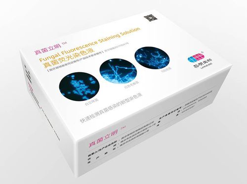
The FungusClearTM Fungal fluorescence staining solution is used for the rapid Identification of various fungal infections in human fresh or frozen clinical specimens, paraffin or glycol methacrylate embedded tissues. Typical specimens include scraping, nail and hair of dermatophytosis such as tinea cruris, tinea manus and pedis, tinea unguium, tinea capitis, tinea versicolor. Also include sputum, bronchoalveolar lavage(BAL), bronchial wash, and tissue biopsies from invasive fungal infection patients.
INTRODUCTION
Fungi are eukaryotic organisms. Beta-linked polysaccharides are found in fungi cell walls of various organisms such as chitin and cellulose. Various fungal and yeast types will stain fluorescently including Microsporum sp., Epidermophyton sp., Trichophuton sp., Candidia sp., Histoplasma sp. and Aspergillus sp. among others. The kit will also stain Pneumocystis carinii cysts, parasites such as Plasmodium sp., and regions of fungal hyphae undergoing differentiation. Keratin, collagen, and elastin fibers are also stained and may provide structural guidelines for diagnosis.
PRINCIPLE
Calcofluor White Stain is a non-specific fluorochrome that binds with cellulose and chitin contained in the cell walls of fungi and other organisms.
Evans blue present in the stain act as a counterstain and diminishes background fluorescence of tissues and cells when using blue light excitation.
10% potassium hydroxide are include in the solution for better visualization of fungal elements.
A range of 320 to 340 nm can be taken for emission wave lenght and the excitation occurs around 355nm.
Catalogs
No catalogs are available for this product.
See all of Nanjing Liming Bio-products Co., Ltd.‘s catalogsOther Nanjing Liming Bio-products Co., Ltd. products
Others
Related Searches
- Assay kit
- Solution reagent kit
- Blood rapid diagnostic test
- Diagnostic reagent kit
- Rapid lateral flow test
- Immunoassay rapid diagnostic test
- Cassette rapid diagnostic test
- Molecular test kit
- Virus rapid diagnostic test
- Serum rapid diagnostic test
- Respiratory infection test kit
- Plasma rapid diagnostic test
- Histology reagent kit
- Infectious disease rapid diagnostic test
- Whole blood rapid diagnostic test
- COVID-19 detection kit
- Rapid respiratory infection test
- Real-time PCR test kit
- Urine rapid screening test
- Clinical reagent kit
*Prices are pre-tax. They exclude delivery charges and customs duties and do not include additional charges for installation or activation options. Prices are indicative only and may vary by country, with changes to the cost of raw materials and exchange rates.


